Download
1 / 35
360 likes | 465 Views
Chapter 11 CARDIOVASCULAR SYSTEM. The heart is a muscle that pumps blood. Blood vessels carry the blood. PRACTITIONERS. Cardiology (cardi/o = heart) Cardiologist Interventional cardiologist Performs procedures and inserts devices Cardiac surgeon

E N D
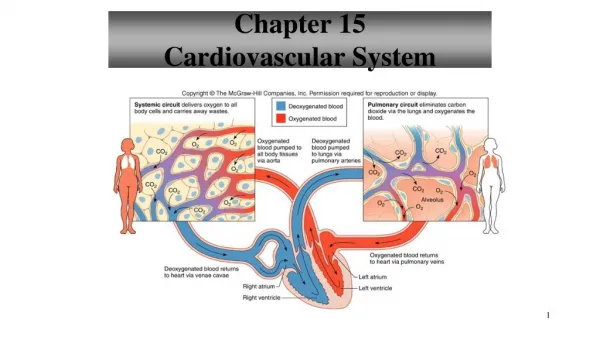
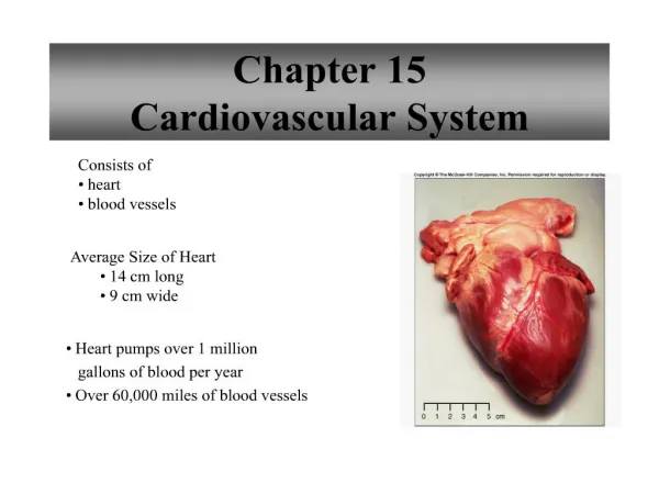
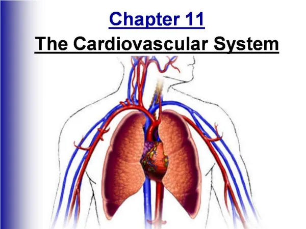
Chapter 11CARDIOVASCULAR SYSTEM The heart is a muscle that pumps blood. Blood vessels carry the blood.
PRACTITIONERS • Cardiology (cardi/o = heart) • Cardiologist • Interventional cardiologist • Performs procedures and inserts devices • Cardiac surgeon • Cardiovascular surgeon (vascul/o = vessel) • Treats vessels
HEART FACTS • Located in center of thoracic cavity • Between the lungs & behind the sternum • Size of your fist • Points (bottom, apex) left • Three-layered wall • Pericardium – outer layer of fibrous tissue • Myocardium – middle layer of thick muscle (my/o) • Endocardium – inner layer of epithelial cells
HEART ANATOMY • Septum (sept/o = partition) • Separates the two sides of the heart (L & R) • 4 chambers • Atria (atrium) – 2 upper chambers (atri/o) • Ventricles – 2 lower chambers (ventricul/o) • Valves connect upper and lower chambers • Bicuspid valve – left side (bi = two) • Also called mitral valve • Tricuspid valve – right side • Open and close together • Prevent blood from flowing back into the atria
HEART ANATOMY (con’t) • 2 additional valves in ventricles • Pulmonary valve –opening of pulmonary artery • Aortic valve –opening of the aorta • Work at the same time
Name the valves (#5 is not a valve)
1. Pulmonary Valve2. Tricuspid Valve3. Mitral (Bicuspid) Valve4. Aortic Valve5. Heart Apex
HEART ACTION • Heart contracts to pump blood • Systole – contraction phase • Diastole – relaxation phase • Heartbeat consists of: • Contraction by atria, then ventricles • Together = cardiac cycle • Average heart beat is 72/min • Cardiac output = volume of blood pumped in 1 min.
- controlled by the cardiac center within the medulla oblongata. The cardiac center signals heart to increase or decrease its rate according to many factors that the brain constantly monitors. Regulation of Cardiac Cycle • Muscle Activity • Body Temperature • Blood ion levels (potassium & calcium)
BLOOD FLOW THROUGH THE HEART • Pulmonary circulation • Right side of heart carries blood to lungs • Return of de-oxygenated blood from the body • Blood flow to pick up oxygenated blood in lungs • Systemic circulation • Left side of heart carries blood to body • Oxygenated blood returns from lungs • Oxygenated blood sent out to body cells via aorta
CONDUCTION SYSTEM • Works like an electrical circuit • Signal flows from • SA node –begins process • Pacemaker of heart • Determines rhythm • AV node • Right and left bundle branches (Bundle of His) • Purkinje fibers
Parts of the Blood • Plasma (55% of the blood) • A straw-colored fluid which is about 90% water and 10% dissolved gases, salts, nutrients, enzymes, hormones, waste products and plasma proteins • Cells (45% of the blood) • RBC’s • Most numerous (5-6 million) • Contain hemoglobin, which is the iron-containing protein that binds oxygen • WBC’s (leukocytes) (4,000-10,000) • They are the army of the circulatory system • May increase dramatically when the body is fighting an infection • Platelets (250,000 – 400,000) • Help in blood clotting by clumping together at the injury to prevent blood from flowing out of the cut
BLOOD VESSELS • Vessel • Vas/o • Angi/o • Arteries (arteri/o) • Carries blood away from the heart • Largest artery is the aorta • Get smaller – form arterioles (arteriol/o) • Arterioles change into capillaries • Nutrients and gases are exchanged
VESSELS (con ‘t) • Venous system • Carries blood back to heart • Capillaries form into tiny venules • Veins (ven/o , phleb/o) • Largest = superior and inferior venae cavae • Veins have one-way valves to prevent back flow of blood • Lumen: central opening of vessel • Pulse: artery expanding and contracting • Arterial system helps maintain body’s blood pressure: constrict and dilate to keep an even pressure gradient
HEART FACTS • Heart has a circulatory system to nourish the heart muscle • Called coronary circulation (coron/o = circle or crown) • Right and left coronary arteries • Decreased blood flow leads to heart tissue death • Myocardial infarction (heart attack) • Blood pressure (BP) • Hypertension = Elevated BP • Increased resistance in vessels – Poor blood flow • Hypotension • Too little blood flowing to organs • Read: systolic pressure (#) /diastolic pressure (#) • Goal is ~ 120/80
WELLNESS & ILLNESS • Listen to the heart with a stethoscope • Lub - dub = S1, S2 • S1 – closing of AV valves as ventricles contract (systole) • S2 – closing of semilunar valves closing in relaxation diastole • Listen to specific areas of heart for: • Abnormal heart sounds • Valve defects
Heart Sounds - Opening and Closing of Valves, "Lub Dub" Stethoscope - instrument to listen and measure heart sounds
FETUSES & NEWBORNS • At birth, lungs inflate and aeration begins • Congenital heart defects • Impaired cardiac function • ASD: opening between atrium • VSD: opening between ventricles • PDA: persistent fetal circulation • Coarctation of aorta: narrowing of descending portion of aorta
CHILDREN • Murmurs • Innocent murmurs • Functional murmurs
ADULTS –Risk Factors • Risk factors for heart disease • Cannot be controlled: • Age, family history, gender • Can be modified/treated/controlled: • Smoking • Physical Activity • Body mass Index: keep less than 25 (weight) • Blood pressure: keep less than 120/80 • Blood glucose: keep less than 140 (random) • Cholesterol • Keep total below 200 • HDL, LDL, triglyceride levels
ADULTS • PROBLEMS • Arteriosclerosis (scler/o = hardening) • Affects walls of small vessels • Angina pectoris • Severe pain in chest (and may radiate) • Decreased blood flow to heart • Abnormalities in vessels • Aneurysm • Ballonlike swelling of an artery • Atherosclerosis (ather/o = pasty material) • Blockage caused by lipid deposits
SENIORS • Aging changes • Elevated BP • Normal calcification of vessels • Coronary heart disease • General term • Congestive heart Failure • Poor pumping ability of heart – poor body perfusion • Arrhythmias (rhythm = beat) • Irregular heart rhythm • Ectopic beats (ecto = outer) • Heartbeat outside regular rate and rhythm • Outside SA node regulation
GENERAL TERMS • Bradycardia (brady = slow) • Tachycardia (tachy = rapid) • Cardiomegaly (megal/o = enlargement) • Vasoconstriction • Narrowing of a vessel • Vasodilation • Widening of a vessel • Varicose Vein • Dilated and twisted veins, usually in legs
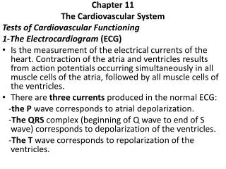
TESTS • Blood testing − lipid risk panel • Electrocardiogram (ECG) • Ultrasound of heart • EKG • Tracing electrical activity of the heart • Identify heart muscle changes
An EKG is printed on paper covered with a grid of squares. Notice that five small squares on the paper form a larger square. The width of a single small square on EKG paper represents 0.04 seconds. - A common length of an EKG printout is 6 seconds; this is known as a "six second strip." Interpreting EKGs
HEART RHYTHMS • EKG tracings
PROCEDURES • Interventional cardiology • Cardiac catheterization (angiogram) • To determine flow of blood through heart and main vessels • Use catheter and dye • Balloon Angioplasty (PTCA) • Balloon catheter inserted into blocked coronary artery, then inflated to push plaque against vessel walls • Endarterectomy • Removal of plaque from an artery • Common in carotids • Coronary bypass graft (CABG) • Cardiac vessels replaced with healthy ones
DRUGS • Improve function of heart muscle • Beta-blockers, antiarryhythmics, Digoxin • Eliminate access fluid • Diuretics (Lasix) – CHF • Ensure flow of blood through vessels • Anticoagulants • Decrease blood pressure • Antihypertensives • Decrease serum cholesterol levels • Hypolipidemics