Download
1 / 27
270 likes | 360 Views
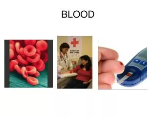
Cardiovascular System: Blood Chapter 11 2013. Blood has several important functions and unique physical characteristics. 1. Transporting dissolved gases, nutrients, hormones and metabolic wastes. 2. Regulating the pH and ion composition of interstitial fluids throughout the body

E N D
Cardiovascular System: Blood Chapter 11 2013
Blood has several important functions and unique physical characteristics. • 1. Transporting dissolved gases, nutrients, hormones and metabolic wastes. • 2. Regulating the pH and ion composition of interstitial fluids throughout the body • 3. Restricting fluid losses at injury sites. • 4. Defending against toxins and pathogens • 5. Stabilizing body temperature.
Blood is made up of plasma and formed elements. • Temperature: roughly 38oC (100.4oF) • Viscosity: 5X more viscous as water = more sticky, more cohesive and resistant to flow. • pH: slightly alkaline with pH between 7.35 and 7.45
Plasma, contains significant quantities of plasma proteins. • 3 main types • Albumin = majority. Maintains osmotic pressure of plasma • Globulins: include antibodies and transport proteins • Fibrinogen: functions in blood clotting. Liver synthesizes most of the plasma proteins.
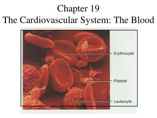
Red Blood Cells • 99% of the formed elements in plasma • Measured by hematocrit or % of whole blood volume occupied by formed elements. • Males have 5.4 million RBC’s / microliter • Females have 4.8 million RBC’s/ microliter
RBC’s Structure • Biconcave disc allows for large surface area for diffusion. • Flexible to squeeze through narrow capillaries • During their formation, RBCs lose most of their organelles including mitochondria • Don’t divide or synthesize any proteins. • Formed in the red bone marrow through erythropoiesis
Erythropoietin (EPO) stimulates the formation of RBC’s • Released by the kidneys when tissues are exposed to low oxygen levels. • EPO is released • During anemia, • When blood flow to the kidneys declines, • When oxygen content of the air in the lungs declines • When the respiratory surfaces of the lungs are damaged.
Hemoglobin • Hb = 95% of an RBCs proteins • Binds oxygen to an iron ion • Iron + oxygen give the blood a bright red color • Dark color when oxygen is not bound. • A low hematocrit, or a reduced oxygen-carrying capacity is a condition called anemia. • Anemia causes muscle fatigue, weakness, and a general lack of energy
Abnormal Hemoglobin • Read about thalassemia and sickle cell anemia on page 385 of your book.
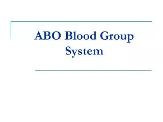
ABO blood types and Rh system are based on antigen-antibody responses. • The presence of absence of specific surface antigen in RBC membranes determines your blood type • There are four blood types.
Hemolytic Disease of the Newborn • Sometimes the Rh factor of the fetus is different than the mother. • If the mother is Rh- and her child is Rh+, there is a chance that during delivery, the fetal and maternal blood may mix, exposing the mother to the Rh antigens of the baby. The mother will begin to produce anti-Rh antibodies. • These antibodies will cross the placenta and attach the RBC’s of the next Rh+ fetus, hemolyzing them.
White blood cells contribute to the body’s defenses • WBC’s = leukocytes • Larger than RBC’s • Have a nucleus and other organelles • Lack hemoglobin. • Defend the body against pathogens, remove toxins, wastes and abnormal or damaged cells.
WBC circulation and movement • Four characteristics • All are capable of amoeboid movement • All can migrate out of bloodstream through process of diapedesis • All are attracted to specific chemical stimuli (positive chemotaxis) • Neutrophils, eosinophils, and monocytes are capable of phagocytosis
Types of WBC’s • Neutrophils: first to arrive at an injury • Attack bacteria by phagocytosis • Short life span (hours) • Forms pus at the wound • Eosinophils • Attack objects coated with antibodies • Increase in numbers during a parasitic infection or allergic reaction • Basophils • Small cells that accumulate in damaged tissues where they release heparin and histamine.
Monocytes • Large cells • Aggressive phagocytes • Release chemicals that draw other WBC’s to injury • Lymphocytes • Migrate between the blood stream and tissues • Do not use phagocytosis • Some secrete antibodies • May be active during a viral infection.
Platelets function during clotting process • One of the formed elements of the blood • Formed as cell fragments from megkaryocytes in red bone marrow • Initiate clotting process and help close injured blood vessels
Hemostasis involves vascular spasm, platelet plug formation, and blood coagulation • Hemostasis is the process that halts bleeding and prevents the loss of blood through the walls of damaged vessels. At the same time it also establishes a framework for tissue repairs.
Phases of Hemostasis • 1. Vascular Phase • Cutting the wall of a blood vessel triggers the contraction of the smooth muscles • Called vascular spasm • Lasts about 30 min. • 2. Platelet Phase • Platelets begin to attach to sticky endothelial surfaces within 15 seconds forming a platelet plug. • 3. Coagulation Phase • Begins at least 30sec after injury • Involves a complex series of steps leading to a blood clot
The Clotting Process • Cannot occur if plasma doesn’t contain clotting factors • During clotting phase these proteins interact in sequence or cascade • Extrinsic pathway begins in the wall of the blood vessel • Intrinsic pathway begins in the blood stream • Common pathway-both process join here through the activation of Factor X
Calcium ions and vitamin K affect almost every aspect of the clotting process. • All three pathways require the presence of calcium ions so any disorder that lowers plasma calcium ion concentration will impair blood clotting • Vitamin K must be present for the liver to synthesize four of the clotting factors so a deficiency of vitamin K inactivates the clotting system.