Download
1 / 46
470 likes | 608 Views
Chapter 11 The Cardiovascular System. Biology 112 Tri-County Technical College Pendleton, SC. Functions of CV System. Major function is transportation

E N D
Chapter 11 The Cardiovascular System Biology 112 Tri-County Technical College Pendleton, SC
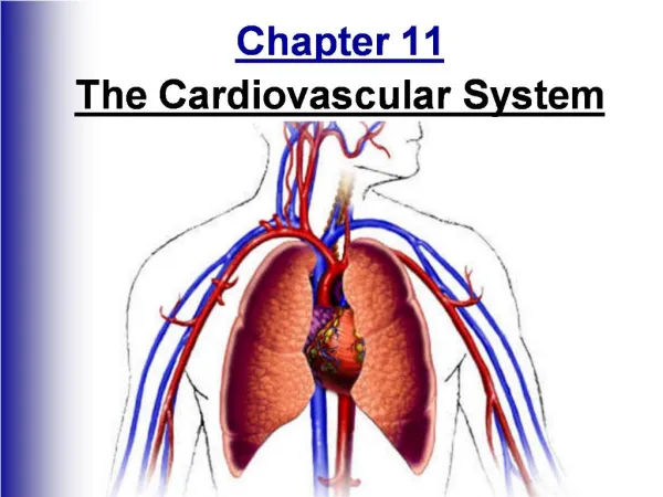
Functions of CV System • Major function is transportation • Using blood as transport vehicle, system carries oxygen, nutrients, cell wastes, hormones, and many other substances vital to homeostasis to and from cells • The force to move blood around the body provided by beating heart • Heart located in body thorax flanked by lungs
Have a heart… • More pointed apex pointed toward left hip & rests on diaphragm • Broader posterosuperior aspect, or base from which great vessels of body merge, points toward right shoulder and lies beneath the second rib
Coverings of the Heart • Enclosed by double sac of serous membane (pericardium) • Thin visceral pericardium (epicardium) hugs external surface of heart and is part of heart wall • Epicardium continuous at heart base with loosely applied parietal pericardium • Serous fluid (slippery lubricating) produced by serous pericardial membranes • Allows heart to beat almost frictionless environment as pericardial layers slide across each other
Walls of the Heart • Epicardium, Myocardium, Endocardium • Epicardium is thin visceral pericardium • Myocardium (middle layer) is composed of thick bundles of cardiac muscle twisted & whorled into ringlike arrangement • Myocardium that actually CONTRACTS • Endocardium (innermost) is thin, glistening sheet of endothelium that lines chambers • Continuous with lining of blood vessels entering and leaving heart
Heart Chambers • Four hollow chambers or cavities • Two atria and two ventricles—all lined with endocardium • Superior atria primarily receiving chambers • Inferior, thick-walled ventricles discharging chambers (actual pumps of heart • Septum dividing heart longitudinally referred to as interatrical and interventricular septum
Great Vessels of the Heart • Superior and inferior vena cavaright atria • Right ventriclepulmonary truckright and left pulmonary arterieslungs • O2 rich blood from lungs to heart4 pulmonary veins (pulmonary circuit)left atrialeft ventricleaortasystemic arteriesbody tissues (systemic circuit)
Heart Valves • Four valves whose general function is to prevent backflow of blood • Atrioventricular (AV) valves between atrial and ventricular chambers on each side • Left AV valve (bicuspid/mitral) has 2 cusps (flaps) & prevents backflow into left artium • Right AV valve (bicuspid) has 3 cusps & prevents backflow into right atrium • Pulmonary semilunar valve—right ventricle • Aortic semilunar valve—left ventricle
Blood Flow • From body into right atrium via vena cava • Right atriumright ventricle via tricuspid • Right ventriclelungs via pulmonary semilunar valve and pulmonary arteries • Lungsleft atrium via pulmonary veins • Left atriumleft ventricle via bicuspid • Left ventriclebody via aortic semilunar valve and aorta
Cardiac Circulation • Blood in heart does NOT nourish myocardium • Right and left coronary arteries responsible • Branch from base of aorta, encircle heart in atrioventricular groove • Anterior interventricular & circumflex arteries on left and posterior interventricular and marginal arteries on right compressed when ventricles contracting and fill when ventricle relax • Myocardium drained by several cardiac veins • Empty into coronary sinus (enlarged vessel on backside of heart • Coronary sinus empties into right atrim
Some Key Terms • Pericarditis-inflammation of pericardium often from decrease in amount of serious fluid • Pericardial layers bind and stick to each other • Forms painful adhesions interfering with heart movements • Valvular Stenosis-valve flaps become stiff (repeated bacterial infection of endocardium) • Forces heart to beat more vigorously than normal • Workload increases, heart weakens, and may fail
Key Terms, cont. • Myocardial infarction (heart attack or coronary)-myocardial cells NOT receiving adequate blood supply • Ischemic heart cells begin to die
Impulse Conduction • 2 types of controlling systems regulate • Nerves of ANS and intrinsic conduction system (nodal system) • ANS acts like “brakes” or “accelerator” depending on which division is activated • Intrinsic system composed of special tissue found no where else in body • Cross between nervous & muscle tissue • Causes depolarization in only ONE direction-from atria to ventricles
Impulse, cont. • Nodal system composed of sinoatrial (SA) node (right atrium); atrioventricular (AV) node(junction of atria & ventricles); AV bundles (bundle of His); Right and Left bundle branches (interventricular septum); and Purkinje fibers (within muscle of ventricular wall) • SA (Pacemaker) fires • Impulse spreads through atria to AV node • Atria contract
Impulse, III • AV node delays impulse preventing simultaneous atria/ventricle contraction • allows complete emptying of atria • Impulse conducted to AV bundle, bundle branches, and Purkinje fibers • Ventricles contract • Heart beats to internal rhythm but body can speed it up or slow it down • Vagus nerve <; Sympathetic fibers >
Heart Block • UNC, depolarization waves reach ventricles only through AV node • Damage to AV node can partially/totally release ventricles from control of SA node • Ventricles begin to beat at their own rate (which is much slower) some or all the time • This condition is called heart block
Three Key Terms • Ischemia is lack of adequate blood supply to heart muscle • May lead to fibrillation (rapid, uncoordinated shuddering of hear muscle) which makes heart useless as pump and major cause of death from heart attacks • Often followed by arrest • Bradycardia is heart rate substantially slower than normal (less than 60 beats per minute) • Tachycardia is rapid heartbeat (over 100 beats per minute) • May progress to fibrillation
Cardiac Cycle • Refers to events of ONE heartbeat • Average is 75 beats per minute so cycle normally about 0.8 seconds • Systole means heart contraction • Diastole means heart relaxation • Atria are in systole at same time and ventricles are in systole at the same time
Heart Sounds • Lub-Dup • First sound (lub) = closing of AV valves • Second sound (dup) caused by closing of semilunar valves at end of systole • Lub sound longer and louder; Dup tends to be short and sharp • Abnormal/unusual sounds called murmurs • Caused by turbulance of blood flow • Fairly common in children/elderly people
Sounds, cont. • Murmurs may indicate valve problem • If valve does NOT close tightly (incompetent), swishing will be heard as blood flows back through partially “open” valve • Distinct sounds also heard when blood flows turbulently through “stenosed” (narrowed) valves
Cardiac Output • CO is amount of blood pumped out by each side of heart • Actually each ventricle in ONE minute • Stroke volume is volume of blood pumped out by each ventricle with each heartbeat • SV >s as force of ventricular contraction >s • CO is PRODUCT of heart rate (HR) x stroke volume (SV); CO=HR x SV
Output, cont. • 75 beats/min x stroke volume of 70 ml/beat equals 5250 ml/min • Normal blood volume = ~5000 ml, entire blood supply passes through heart once each minute • SV regulated by many factors • > volume or speed of venous return >s SV & force of contraction • < volume or speed of venous return <s SV & force of contraction
Output, cont. • “Starlings law of the heart: more heart muscle is stretched, the stronger contraction • Critical factor “stretching” heart muscle is venous return • Insures volume of blood going out equals volume of blood coming in (vice-versa) • REGULATION of heart rate dependent of many factors
Output and Regulation, cont. • Heart rate can be changed temporarily by autonomic nerves • Sympathetic nerves stimulate SA and AV nodes and heart muscle itself to > rate • Parasympathetic nerves (vagus) slow and steady heart giving rest time during noncrisis • Epinephrine and thyroxine >s heart rate
Output & Regulation, cont. • Physical factors (age, gender, exercise, and body temperature) influence heart rate • Hypocalcemia (reduced levels of ionic calcium) depress the heart • Hypercalcemia (> levels of ionic calcium) causes prolonged contraction to point heart may stop entirely • Hypokalemia (reduced levels of ionic potassium causes feeble heart beat and abnormal rhythms appear • heatheart rate and coldheart rate
Congestive Heart Failure • Pumping efficiency depressed so circulation inadequate to meet tissue needs = congestive heart failure • One side can fail independently of the other • Left side fails = Pulmonary congestion occurs • vessels in lungs swells-fluid leaks-pulmonary edema-if untreated, person suffocates • Right side fails = Peripheral congestion occurs • blood backs up in systemic circulation-edema most noticeable in distal body parts (swollen feet, ankles, fingers) • Failure of one side puts strain on other side & eventually whole heart fails
Electrocardiogram (ECG) • Impulses cause electrical currents to pass through body • Can be detected by electrocardiograph • Three recognizable waves (P, QRS complex, & T) • P wave first and small-depolarization of atria before they contract • QRS complex is large wave-depolarization of ventricles; precedes their contraction • T-wave results repolarization of ventricles • Atrial repolarization normally hidden by QRS • May reveal heart problems: abnormal waves; changes in timing; fibrillation
Arterial Pulse • Expansion and recoil of artery occurring with each ventricle beat creates PULSE • Pulse rate = heart rate • Averages 70-80 per minute • Influenced by activity, postural changes, emotions • May be taken at any artery close to surface • Temporal, carotid, brachial, & radial most common
Blood Pressure… • Is pressure blood exerts against inner walls of blood vessels • Force that keeps blood circulating • USD, understood to mean pressure within large systemic arteries NEAR heart • Pressure highest in large arteries and <s thru pathway • Reaches 0 or negative at vena cava • Return dependent on valves in larger veins, milking activity of skeletal muscles, and pressure changes in thorax
Auscultatory Method for BP • Systemic arterial BP measured indirectly • System uses brachial artery of arm • Systolic pressure=pressure at peak of ventricular contraction • Diastolic pressure=pressure when ventricles are relaxed • “Normal” is 120/80 but range is 110-140/75-80 • Varies with age, weight, mood, race, activity, and posture
BP Math…or whatever • BP = cardiac output x peripheral resistance • Cardiac output increases, BP increases • Peripheral resistance increases; BP increases • Cardiac output decreases; BP decreases • Peripheral resistance decreases; BP decreases
Factoring the factors • Sympathetic division of ANS causes vasoconstriction >ing peripheral resistance which >s BP • Parasympathetic division of ANS causes vasodilation <ing peripheral resistance which <s BP • Kidneys help regulate BP by altering blood volume • Retain or excrete water • Also produce RENIN
Factors, cont. • If BP low, kidney cells release enzyme renin into blood • Renin triggers cascade of reactions that produce Angiotensin II which is potent vasoconstrictor • Vasoconstriction raises BP • AT II stimulates adrenal cortex to release Aldosterone (hormone that >s sodium ion reabsorption by kidneys • Water follows sodium ions thus blood volume and BP both rise
Enough of factoring, already.. • Epinephrine >s heart rate and BP • Nicotine >s BP by causing vasoconstriction • Alcohol/Histamine < BP by causing vasodilation • Diuretics cause kidneys to excrete more water which reduces blood volume thereby lowering BP • TOO much salthypertonic blood which absorbs water from tissues increasing blood volume and BP
The end of factoring…Yeah!! • TOO much fatsexcessive fats in blood • Makes blood more viscous and more difficult to pump • Peripheral resistance is increased which increases the blood pressure
Hypotension • Low BP = systolic < 100 mm Hg • Physical conditioning and/or health • Orthostatic hypotension = temporary drop in BP resulting in dizziness upon rising • May be slow reacting sympathetic NS and blood pooling in lower limbs reducing BP and blood delivery to brain • Chronic hypotension may be result of inadequate blood proteins, low viscosity, and/or low pressure
Hypertension • Sustained elevated arterial pressure of 140/90 or higher • Heart has to work against increased resistance and has to work HARDER • Myocardium enlarges; when strained beyond capacity to respond, weakens and becomes flabby • Causes small tears in endothelium of blood vessels that > progress of artherosclerosis
Hypertension, cont. • Most cases (90%+) are primary or essential which CANNOT be accounted for by any specific organic cause • Diet, obesity, heredity, race, stress involved • Treatment for hypertension • diuretics • vasodilators/beta blockers • ACE-inhibitors (inhibit renin) • Cardiac inhibitors (calcium channel blockers) • Lifestyle changes