Download
1 / 37
370 likes | 839 Views
Anatomy and function of the spinal cord and nerves. Spinal cord Ch. 14, 17. Spinal cord is protected by vertebrae, meninges and CSF. Canal formed by foramina of vertebrae Meninges Dura mater Arachnoid mater Pia mater CSF formed in brain Found in subarachnoid space.

E N D
Anatomy and function of the spinal cord and nerves Spinal cord Ch. 14, 17
Spinal cord is protected by vertebrae, meninges and CSF • Canal formed by foramina of vertebrae • Meninges • Dura mater • Arachnoid mater • Pia mater • CSF formed in brain • Found in subarachnoid space
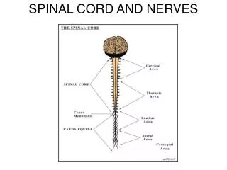
31 pairs of spinal nerves • 8 cervical • 12 thoracic • 5 lumbar • 5 sacral • 1 coccygeal • All are mixed nerves
Internal structure of spinal cord • Roots connect spinal nerves to spinal cord • Dorsal root- sensory neurons • Ganglion-cell bodies of sensory neurons • Ventral root- motor neurons • Horns- gray matter; integrates sensory and motor information • Anterior, posterior, lateral • White matter- myelinated; sensory and motor tracts • Columns- anterior, posterior, lateral
Regions of white and gray matter • Dorsal (posterior) horns- sensory neurons • Ventral (anterior) horns- motor neurons • Lateral horns- autonomic (motor) neurons • Columns- ascending (sensory) and descending (motor) tracts
Sensory and motor tracts • Named according to its position and pathway • Sensory impulses move up tracts and columns • Join the “pool” of sensory information • Voluntary motor output emanates from cerebral cortex (direct pathways) • Involuntary output originates in brain stem and hypothalamus (indirect pathways)
Spinal nerves • Comprise the peripheral nervous system (PNS) • Connect the CNS to muscles, sensory receptors and glands • 31 pairs of spinal nerves • Posterior and anterior roots (mixed nerves)
Distribution of spinal nerves • Branches (rami) • Posterior (muscles and surfaces; anterior) • Meningeal branch (CNS) • Rami communicantes- autonomic system • Plexuses- network of axons • Cervical, brachial, lumbar, sacral
“Map” of spinal nerves Motor fibers Sensory fibers
Major plexuses example: cervical plexus • Origin (C1- C5) • Superficial or deep • Superficial- skin; mostly sensory • Deep-muscle; largely motor • Severing spine above phrenic nerve causes respiratory arrest
More plexuses • Brachial • Shoulders, upper limbs • Axial, musculocutaneous, radial, medial, ulnar • Lumbar • Anterolateral abdominal wall, genitalia, part of lower limbs • Sacral and coccygeal • Buttocks, perineum, lower limbs • Damage can cause loss of sensation, palsy, loss of motor control • Intercostal nerves directly innervate muscles (no plexuses)
Dermatomes • Sensory neurons convey information form skin to CNS • Dermatomes- areas of skin that provide sensory input to a particular pair of nerves • Some overlap • Can be useful for diagnosis
Reflex arcs may be somatic or autonomic • Patellar reflex (knee-jerk) • Tap patellar ligament; quadriceps femoris contracts • Achilles reflex (ankle-jerk) • Tap calcaneal tendon; gastrocnemius and soleus muscles contract • Babinski sign- test for continuity of the corticospinal tract (goes from positive to negative with development) • Abdominal reflex • Can be useful for diagnosis of chronic disease or nerve damage
Somatic senses (pain, thermal, tactile, proprioreceptive) Somatic motor neurons- skeletal muscle Usually voluntary Motor units Acetylcholine Autonomic sensory (chemoreceptors, mechano- (stretch) receptors Motor neurons regulate visceral activities; can function independently of ANS Preganglionic and postganglionic neurons Acetylcholine or norepinephrine Autonomic vs. somatic nervous system
Different types of ganglia • Sympathetic • Sympathetic trunk • Innervate organs above the diaphragm • Superior, middle, inferior cervical ganglia • Prevertebral • Below the diaphragm • Celiac, superior and inferior mesenteric ganglia • Parasympathetic • Preganglionic axons are longer than in sympathetic ganglia • Preganglionic neurons are myelinated, postganglionic neurons are not
Sympathetic (thoracolumbar) • Sympathetic chain ganglia, paired • Head and thoracic cavity • Collateral ganglia, unpaired • Abdominopelvic cavity • Suprarenal medullae, paired • hormonal
Functions of the sympathetic division • Sympathetic chain ganglia • Diversion of blood flow to skeletal muscles and brain • Sweating; pupil dilation • Collateral ganglia • Vasoconstriction; restrict urinary function • Suprarenal medullae • Release of epinephrine and norepinephrine
Parasympathetic system • Ganglia are located near, or in, target organs • Effects are directed more toward specific target organs
Regulation by dual innervation • Systems intersect in plexuses • Cardiac • Pulmonary • Esophageal • Celiac • Inferior mesenteric • Hypogastric
Postganglionic neurons • Sympathetic system • One presynaptic neuron can diverge into many • Many organs can be affected at once (divergent) • May extend to adrenal medullae • Parasympathetic system • Many presynaptic neurons can converge on a single effector • Effect can be localized to a single effector
Action by neurotransmitters in ANS • Acetylcholine (cholinergic neurons) • All preganglionic • Sympathetic postganglionic innervation of sweat glands • All parasympathetic postganglionic neurons • Nicotinic, muscarinic receptors • Norepinephrine (adrenergic) • α and β receptors • Can be excitatory or inhibitory
Functions of ANS • “fight or flight” (sympathetic) • More widespread and longer lasting • Norepinephrine and epinephrine can act as hormones as well as neurotransmitters • “rest and digest” (parasympathetic) • Salivation, lacrimation, urination, digestion and defecation
Control of autonomic functions • Reflexes • Blood pressure • Digestion • Defecation and urination • Control within brain • Brain stem (cardiovascular, swallowing , digestion) • Spinal cord (elimination) • Control and integration center is hypothalamus
Sympathetic vs parasympathetic effects • Parasympathetic tends to be organ-specific • More reflexes • Sympathetic tends to be more holistic • Divergence • Hormonal distribution