Download
1 / 19
250 likes | 313 Views
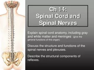
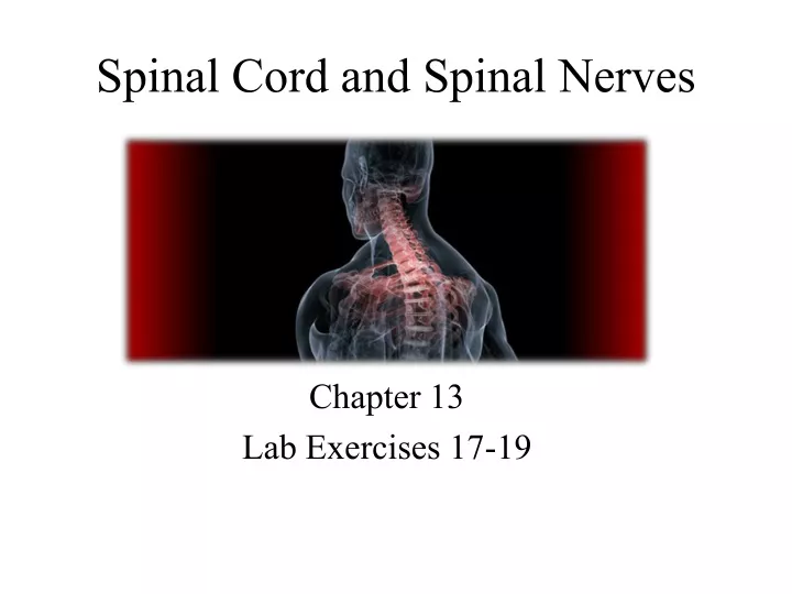
Spinal Cord and Spinal Nerves. Chapter 13 Lab Exercises 17-19. Introduction. The purpose of this part of the course is to: Identify & describe anatomical features of spinal cord & spinal nerves Discuss functions of spinal cord & spinal nerves homeostasis Understand spinal reflex arcs.

E N D
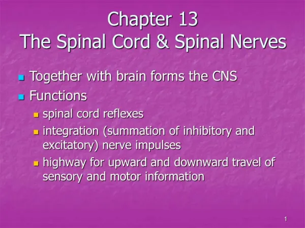
Spinal Cord and Spinal Nerves Chapter 13 Lab Exercises 17-19
Introduction The purpose of this part of the course is to: Identify & describe anatomical features of spinal cord & spinal nerves Discuss functions of spinal cord & spinal nerves homeostasis Understand spinal reflex arcs Integration of EPSP + IPSP Sensory and motor tracts
Functions of the Spinal Cord • Processes reflexes • Integrates nerve impulses • Conducts sensory impulses to the brain and motor impulses to effectors
Protection of the Spinal Cord The spinal cord is protected by: • Bone • Meninges • CSF
Meninges The meninges are composed of three layers • Dura mater • Arachnoid mater • Pia mater • Denticulate ligaments 3 2 1
3 Meninges Epidural space Subdural space Subarachnoid space A spinal tap can be done to withdraw CSF for diagnostics 2 1
SUPERIOR Fourth ventricle Cerebellum of brain (cut) Glossopharyngeal (IX) and vagus (X) nerves Occipital bone (cut) Accessory (XI) nerve Posterior median sulcus Gracile fasciculus Vertebral artery Cuneate fasciculus Denticulate ligament Posterior (dorsal) rootlets of spinal nerve Dura mater and arachnoid mater (cut) INFERIOR (b) Posterior view of cervical region of spinal cord
View Transverse plane Dura mater and arachnoid mater POSTERIOR Spinal cord Pia mater Spinous process of vertebra Epidural space Subarachnoid space Superior articular facet of vertebra Posterior (dorsal) root of spinal nerve Posterior (dorsal) ramus of spinal nerve Denticulate ligament Anterior (ventral) root of spinal nerve Spinal nerve Anterior (ventral) ramus of spinal nerve Transverse foramen Body of vertebra Vertebral artery in transverse foramen ANTERIOR Figure 13.1 (b) Transverse section of the spinal cord within a cervical vertebra
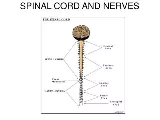
External Anatomy of the Spinal Cord Begins… Ends… Landmarks
Fig. 13.2 External Anatomy of Spinal Cord Medulla oblongata C1 CERVICAL PLEXUS (C1–C5): C2 Lesser occipital nerve C3 Atlas (first cervical vertebra) Great auricular C4 C5 Ansa cervicalis CERVICAL NERVES (8 pairs) C6 C7 Transverse cervical nerve Cervical enlargement C8 Supraclavicular nerve T1 T2 Phrenic nerve First thoracic vertebra T3 BRACHIAL PLEXUS (C5–T1): T4 T5 Musculocutaneous nerve THORACIC NERVES (12 pairs) T6 Axillary nerve T7 Median nerve T8 Lumbar enlargement Radial nerve T9 Ulnar nerve T10 First lumbar vertebra Intercostal (thoracic) nerves T11 Subcostal nerve (intercostal nerve 12) Conus medullaris T12 LUMBAR PLEXUS (L1–L4): LUMBAR NERVES (5 pairs) L1 Iliohypogastric nerve Cauda equina L2 Ilioinguinal nerve Genitofemoral nerve L3 Ilium of hip bone Lateral femoral cutaneous nerve L4 Sacrum Femoral nerve L5 Obturator nerve SACRAL NERVES (5 pairs) S1 SACRAL PLEXUS (L4–S4): S2 COCCYGEAL NERVES (1 pair) S3 Superior gluteal nerve S4 Inferior gluteal nerve S5 Filum terminale Sciatic nerve: Common fibular nerve Tibial nerve Posterior cutaneous nerve of thigh Posterior view of entire spinal cord and portions of spinal nerves Pudendal nerve
Internal Anatomy of the Spinal Cord Gray matter White matter Landmarks
Cervical (Segment C6) Table 13.1 Spinal Cord Segments Thoracic (Segment T5) Lumbar (Segment L4) Sacral (Segment S3)