Download
1 / 22
240 likes | 572 Views
Anatomy of Spinal Nerves. The Peripheral Nervous System. Introduction: PNS – all neural structures outside the brain and spinal cord. Provides links to and from the external environment . Peripheral Nervous System. Structural division = cranial nerves & spinal nerves.

E N D
The Peripheral Nervous System Introduction: • PNS – all neural structures outside the brain and spinal cord. • Provides links to and from the external environment.
Peripheral Nervous System Structural division = cranial nerves & spinal nerves. Functional divisions = Afferent (sensory) & Efferent (motor).
The Cranial Nerves • 12 pairs of nerves – first two arise from forebrain and remaining 10 arise from the brain stem • Numbered 1 to 12 from anterior to posterior • Names indicate primary functions or areas served • Some are mixed nerves; some are purely sensory; some are purely motor (although motor nerves may carry some afferent fibres from proprioceptors)
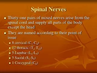
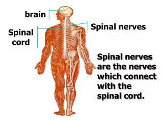
Spinal Nerves • 31 pairs of nerves coming off the spinal cord. • They are all mixed nerves – i.e. containboth afferent (sensory) and efferent (motor) fibres. • Supply all parts of the body except for the head and some areas of the neck. • Named according to their point of emergence from the spinal cord.
Mixed Spinal Nerves Structurally, a spinal nerve attaches to the spinal cord via: • A Ventral Root – which attaches to the anterior horns of grey matter & • A Dorsal Root – which attaches to the posterior horns of grey matter. Note: The dorsal root is characterised a swelling (the Dorsal Root Ganglion)
Mixed Spinal Nerves Functionally: Ventral root – carries sensory input (messages in) Dorsal Root – carries motor output (messages out)
Distribution of Spinal Nerves Spinal nerves branch into several rami. • Dorsal Ramus – supplies the skin and deep muscles of the back • Ventral Ramus – supplies superficial back muscles, lateral & anterior trunk, limbs • Rami Communicantes – serve visceral organs (part of Autonomic system)
Spinal Nerve Plexuses All ventral rami except T2-T12 form interlacing nerve networks called plexuses. (There are 4 nerve Plexuses). This is achieved by small branches joining with those of adjacent nerves Each resulting nerve of a plexus contains fibers from several spinal nerves. Damage to one spinal segment cannot completely paralyze a muscle.
Reflexes • Reflex – a rapid, unconscious response to a stimulus. examples - stretch reflex; flexor (withdrawal) reflex • A Reflex Arc is a simple neural pathway by which sensory impulses from receptors cause a response in effectors without necessarily travelling to the brain
Components of a Reflex Arc • Receptor – responds to stimulus • Sensory Neuron – transmits message to CNS • Integration Centre (within CNS) – link between sensory and motor neurons • Motor Neuron – impulse transmitted from CNS to effector • Effector – muscle or gland which responds to the nerve impulse
Characteristics of reflexes • Somatic reflex – activates skeletal muscle • Autonomic reflex – activates visceral muscle • Note: Although inborn; most reflexes are subject to modification through learning & conscious effort • Primary Function = protection
Reflex Arc Spinal cord (in cross-section) Stimulus Integration center 3 Sensory neuron 2 Receptor 1 Motor neuron 4 Interneuron Effector 5 Skin Figure 13.1