Download
1 / 31
310 likes | 474 Views
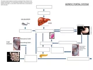
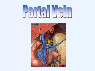
Portal hepatic vein. Foetal circulation. Detailed Anatomy, continued … Vascular Structures in Liver 1. Largest vessels are portal vein and IVC a. Portal Vein : 1. appears on T.S. as tubular, echolucent structure

E N D
Detailed Anatomy, continued … Vascular Structures in Liver 1. Largest vessels are portal vein and IVC a. Portal Vein: 1. appears on T.S. as tubular, echolucent structure 2. courses horizontally from porta hepatis
Detailed Anatomy, continued … 3. walls echogenic due to structures in portal triad b. Left Portal Vein: 1. has more variable course 2. May be difficult to trace on transverse scans
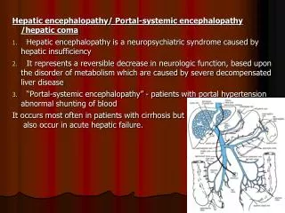
Hepatic Vessels • IVC • Right Portal Vein • Left Portal Vein • Main Portal Vein
Vascular Structures, continued … c. Right Portal Vein: 1. anatomical landmark 2. extends into right lobe 3. branches after porta hepatis 4. L.S. shows “dumbbell” or circular structure with echogenic “collar”
Vascular Structures, continued … d. IVC: 1. To right of aorta 2. Appears to pass through liver 3. Diameter enlarges after renal veins join (~L-1)
Vascular structures, continued … 2. Hepatic Veins: a. Tubular structures b. Enlarge cephalad c. In superior half of liver d. Angles of hepatic vein branches oriented toward IVC e. Walls not echogenic
Hepatic Veins R. Hepatic Vein Middle Hepatic Vein L. Hepatic Vein
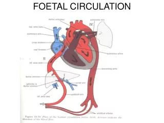
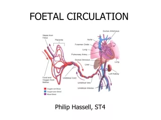
Fetal Circulation • Embryonic lungs and digestive tract nonfunctional • Respiratory functions and nutrition provided by placenta
Placental Blood Supply Figure 21-33a
Placental Blood Supply • Blood flows to the placenta: • through a pair of umbilical arteries • which arise from internal iliac arteries • and enter umbilical cord
Placental Blood Return • Blood returns from placenta: • in a single umbilical vein • which drains into ductus venosus • Ductus venosus: • empties into inferior vena cava
The Neonatal Heart Figure 21-33b
Before Birth • Fetal lungs are collapsed • O2 provided by placental circulation
At Birth • Newborn breathes air • Lungs expand • Pulmonary circulation provides O2
2 Fetal Pulmonary Circulation Bypasses • Foramen ovale: • interatrial opening • covered by valve-like flap • directs blood from right to left atrium • Ductus arteriosus: • short vessel • connects pulmonary and aortic trunks
Cardiovascular Changes at Birth • Pulmonary vessels expand • Reduced resistance allows blood flow • Rising O2 causes ductus arteriosus constriction • Rising left atrium pressure closes foramen ovale
Congenital Cardiovascular Problems Figure 21-34
Congenital Cardiovascular Problems • Develop if proper circulatory changes do not occur at birth