Download
1 / 66
660 likes | 668 Views
Foetal membranes. Fertilization & Implantation. Implantation. Formation of lacunae. Radial arrangement of lacunae. Primary villi. Secondary villi. Tertiary villi. Cytotrophoblastic shell. Maternal surface. Free villi. Term Pacenta. Maternal Cotyledons. Formation of Umbilical Cord.

E N D
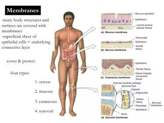
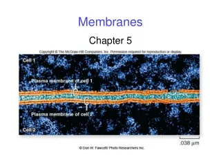
Foetal membranes & Placenta • Structures that are developed from Zygote but do not form the parts of the embryo or foetus • Exception: parts such as yolk sac and allantois • Responsible for protection and nourishment of the embryo and foetus • Includes yolk sac, allantois, amnion and chorion
Foetal membranes • Yolk sac • Development of primitive yolk sac – 2nd week (early part) • Development of secondary yolk sac – 2nd week (Late part) • Covered by extra embryonic splanchnopleuric mesoderm • Lined by endodermal cells • Incorporation into the body of the embryo • Formation of foregut, mid gut and hindgut
Foetal membranes • Yolk sac (contd) • Development of the vitelline duct - disappears • If persists - Meckel’s diverticulum • Vitelline vessels develop in the mesoderm covering the yolk sac • Plays important role in the transfer of nutrients
Foetal membranes • Allantois • 2nd week of IUL • Arises as diverticulum from the caudal end of the yolk sac • Grows into the body stalk • Umbilical vessels develop in close association • Becomes a fibrous cord called urachus • Related to the development of the urinary bladder
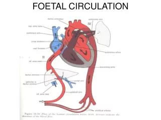
Foetal membranes • Formation of primitive Umbilical cord • Fusion of the mesoderm of the connecting stalk and yolk sac • Covered by the epithelium of the amnion • Primitive umbilical ring - meeting point of the amnion and ectoderm • Attachments • Center of the foetal surface of the placenta • Center of the anterior abdominal wall
Foetal membranes • Structure passing through the umbilical ring • Connecting stalk • 2 umbilical arteries • 1 umbilical vein • Yolksac with vitelline vessels
Foetal membranes-Umbilical cord • When fully developed • Length - 18 inches • Thickness – 1 inch • Cord is twisted presenting false knots • Rarely presents true knots – may tighten – causing fetal death due to anoxia • Long cord may occasionally encircle the neck • Contents of the cord • Whaton’s jelly – Mucoid connective tissue • Urachus • Umbilical vein – one • Umbilical arteries – two • Vessels are longer then the cord
Amnion and amniotic cavity • Amnion is formed by amnioblasts • They are derived from epiblasts • Lines the cavity called amniotic cavity • Filled with clear, watery fluid produced by amniotic cells • Embryo is suspended during the early months of pregnancy • The fluid absorb jolts, prevents the adherence of the embryo to the amnion
Amnion and amniotic cavity • Allows foetal movements • The water in the cavity change every three hours • By 5th month foetus swallows fluid • At the end of the pregnancy urine is added to the fluid • The aquatic habitat provided uniform hydrostatic support for the delicate body of the embryo and prevents asymmetrical development • 800 to 1000 ml at 37 weeks
Amnion and amniotic cavity • Hydramnios – Excess of amniotic fluid (1500-2000 ml) • Oligohydramnios – decreased amount of amniotic fluid (less than 400 ml) • Premature rupture of the amniochorionic membrane
Amniotic bands • Tears in amnion – amniotic bands • Bands encircles the parts of the fetus, particularly the limbs and digits • Results in amputations, ring constrictions, and craniofacial abnormalities
Placenta • Human placenta is discoid in shape • Connects foetus to the uterine wall through umbilical cord • Developed from two sources • Decidua basalis • Chorionic frondosum
Placenta • Development of decidua basalis • Endometrial stromal cells are modified during pregnancy – decidual cells • Contain abundant amount of lipids and glycogen • Decidual basalis, parietalis and capsularis • Development of decidual septa – 4th month • Formation of 15 – 20 cotyledons ( Compartments)
Placenta • Development of chorionic frondosum • 2nd week – uteroplacental circulation • 3rd week – tertiary villi formation • 4th week – heart begins to beat • Rapid growth chorionic villi • Formation of outer cytotrophoblastic shell • Villi are anchored by chorionic plate and cytotrophoblastic shell • Rapid expansion of size and complexity opposite to the decidua basalis
Placental barrier • Maternal hormones do not cross • Hormones that cross the placenta • Thyroxine • Synthetic oestrogen (diethylstilbestrol) • Viruses • Rubella virus • Measles • Poliomyelitis virus • Drugs and drug metabolites
Placenta • Full term placenta • Discoid in shape • Diameter 15 – 25 cms • Thickness – 3 cm • Weight – 500 – 600 grams • Surface • Foetal surface • Amnion • Chorionic vessels • Attachment of umbilical cord
Placenta • Maternal surface • Show cotyledons • Decidua basalis • Placental barrier is of haemochorial type • Dividing membrane between maternal and foetal circulation