Download
1 / 20
200 likes | 331 Views
Basal Ganglia. &. Connections. Basal Ganglia Gross Features. Corpus Striatum Caudate Nucleus Lenticular Nucleus Putamen Globus Pallidus Paleostriatum Pallidum

E N D
Basal Ganglia & Connections
Basal Ganglia Gross Features Corpus Striatum Caudate Nucleus Lenticular Nucleus Putamen Globus Pallidus Paleostriatum Pallidum Corpus Amygdaloid Archistriatum
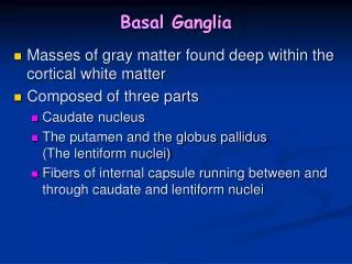
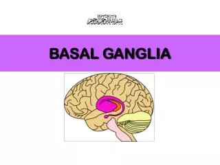
Basal Ganglia Gross Features It is a nuclear mass located within the depths of each cerebral hemisphere. Corpus Striatum Claustrum Amygdaloid body The corpus striatum is the largest component of the basal ganglia. Divided into three components. It includes the caudate nucleus, the putamen and the globus pallidus. The caudate nucleus (CD) is a large C-shaped grey matter located medial to the internal capsule and related throughout its length to the surface of the lateral ventricle.
It has an head that bulges into the lateral wall of the frontal horn of the lateral ventricle. The head tapers to form the body located along the lateral wall of the ventricle. The body in turn becomes further (Continued) attenuated to form the tail which courses along the roof of the temporal horn of the ventricle and terminates at the amygdaloid nucleus. Basal Ganglia Gross Features Amygdaloid N
Basal Ganglia Gross Features • The PU and CD together comprise the neostriatum (neo = new) which is sometimes simply abbreviated striatum. • Histologically, the structure of the CD and PU are similar. • They contain the inhibitory neurotransmitter gamma-amino butyric acid (GABA).
The paleostriatum (paleo = ancient ) or "pallidum" is the forerunner of the mammalian corpus striatum and consists of the globus pallidus (GP) only. The GP is situated medial to the putamen and is separated from it by a thin lamina of myelinated fibers called the external medullary lamina. A similar lamina (medial medullary lamina) divides the GP into a lateral or external division (GPe) and a medial or internal (GPi) division, each of which has its own peculiar set of connections. The GP is traversed by numerous myelinated fibers which give it its characteristic pale appearance in stained sections and from which its name derives. Basal Ganglia Gross Features
Input Portion STRIATUM (Caudate Nucleus and Putamen) Output Portion 1. PALLIDUM (Globus Pallidus) 2. SNr (Substantia Nigra, Pars Reticulata Basal Ganglia connections
Basal Ganglia connections STRIATUM Afferents from 1. Cerebral Cortex [Corticostriatal Projection] 2. Thalamus [Thalamostriatal Projection] 3. SNc (Nigro strial), 5. Amygdaloid Nuclear Complex [Amygdalostriatal Projection] Efferents to 1. Pallidum (GPi & GPe) [Striopallidal Projection] 2. SNr [Strionigral Projection] 3. SNc from striosome
Basal Ganglia Connections Supplementary Motor Area (SMA) STRIATUM (Putamen) Primary Motor Area (M I) pyramidal tract ansa lenticularis THALAMUS (VLo, VApc, CM) PALLIDUM (GPi) lenticular fasciculus LMN
Basal Ganglia Connections Prefrontal Association Cortex STRIATUM (Caudate Nucleus) Primary Motor Area (M I) pyramidal tract THALAMUS (VLm, VAmc, MD) SNr (Substantia Nigra, pars reticulata) LMN
Basal Ganglia (Microcircuitary) Connections glutamate + Motor Cortex Striatum + glutamate glutamate + -- GABA VA-VL complex GP, SNr - GABA
Circuits & Feed Back Cerebral Cortex Striatum Thalamus Pallidum Opp Cer SNr
Organization of the motor system Level 1 Cerebral cortex motor areas 1. The elements Basal Ganglia Level 2 Thalamus 2. Three levels Brain Stem Cerebellum 3. Feedback loops between and within levels. Muscle contraction and Movement Spinal Cord Level 3 Sensory receptors
Disorders Of Basal Ganglia • Hyperkinetic • Chorea, Athetosis & Ballism • Hypokinetic • Akinesia &Bradykinesia
SYDENHAM’S CHOREA Clinical Feature - Complication of Rheumatic Fever - Fine, disorganized , and random movements of extremities, face and tongue - Accompanied by Muscular Hypotonia - Typical exaggeration of associated movements during voluntary activity - Usually recovers spontaneously in 1 to 4 months Principal Pathologic Lesion: Corpus Striatum
HUNTINGTON’S CHOREA Clinical Feature - Predominantly autosomal dominantly inherited chronic fatal disease (Gene: chromosome 4) - Insidious onset: Usually 30-50 - Choreic movements in onset - Frequently associated with emotional disturbances - Ultimately, grotesque gait and sever dysarthria, progressive dementia ensues. Principal Pathologic Lesion: Corpus Striatum (esp. caudate nucleus) and Cerebral Cortex
HEMIBALLISM Clinical Feature - Usually results from CVA (Cerebrovascular Accident) involving subthalamic nucleus - sudden onset - Violent, writhing, involuntary movements of wide excursion confined to one half of the body - The movements are continuous and often exhausting but cease during sleep - Sometimes fatal due to exhaustion - Could be controlled by phenothiazines and stereotaxic surgery Lesion: Subthalamic Nucleus
Parkinson’s Disease PD Disease of mesostriatal dopaminergic system normal Muhammad Ali in Alanta Olympic
Parkinson’s Disease - Paralysis Agitans Substantia Nigra, Pars Compacta (SNc) DOPAminergic Neuron Clinical Feature (1) Slowness of Movement - Difficulty in Initiation and Cessation of Movement
Parkinson’s Disease Paralysis Agitans Clinical Feature (2) Resting Tremor Parkinsonian Posture Rigidity-Cogwheel Rigidity