Download
1 / 17
320 likes | 1.07k Views
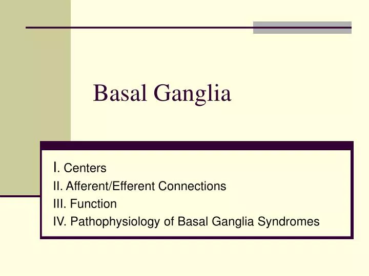
Basal Ganglia. I . Centers II. Afferent/Efferent Connections III. Function IV. Pathophysiology of Basal Ganglia Syndromes. I. General Organization: Centers.

E N D
Basal Ganglia I. Centers II. Afferent/Efferent Connections III. Function IV. Pathophysiology of Basal Ganglia Syndromes
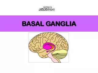
I. General Organization: Centers The basal ganglia (BG) comprise 4 interconnected nuclei that influence behavior by regulating the activities of upper motor neurons. These nuclei are the: • Striatum – Composed of the caudate nucleus and the putamen. • Globus Pallidus (GP) – divided into an internal (GPi) and external (GPe) segments. • Subthalamic Nucleus (STN) • Substantia Nigra (SN) divided into a pars compacta (SNc) and a pars reticulata (SNr).
I. General….(cont.) Striatum. It is derived from the telencephalon. The caudate and putamen are histologically alike and function as a single nucleus called the striatum. In higher mammals, the anterior limb of the internal capsule separates the caudate nucleus from the putamen. • Caudate. Located deep in the cerebral hemisphere, the caudate is a C-shaped nucleus that follows the shape of the lateral ventricle. Its enlarged rostral head forms the lateral wall of the anterior horn of the lateral ventricle. In proceeding caudally, its tapered body is lateral to the body of the lateral ventricle. The body continues as the tail alongside the inferior horn of the lateral ventricle. The tail fuses anteriorly with the amygdaloid nucleus in the medial temporal lobe. • Putamen. Positioned lateral to the head of the caudate and thalamus it does not follow the caudate into the temporal lobe.
I. General……..(cont.) Globus Pallidus. This two-segmented nucleus is positioned medial to the putamen and lateral to both the posterior limb of the internal capsule and thalamus. When viewd together the putamen and globus pallidus form a lens-shaped structure called the “lentiform” nucleus. Sometimes the combination of striatum + globus pallidus is referred to as the “corpus striatum”. Subthalamic Nucleus. Part of the subthalamus, it is an oval-shaped nucleus in the diencephalon, ventral to the thalamus and medial and dorsal to the posterior limb of the internal capsule. It shares reciprocal fiber connections with the globus pallidus. Substantia Nigra. It is located in the rostral midbrain, dorsal to the cerebral peduncles and ventral to the tegmentum (reticular formation). The dorsal part of the nucleus (pars compacta, SNc) is composed of tightly packed dopaminergic (DA) neurons that contain melanin granules. With the naked eye the melanin-containing neurons give this nucleus a black stripe appearance. The ventral part of the nucleus (pars reticulata, SNr) has sparcely distributed gabaergic (GABA) neurons. The SNr and internal globus pallidus (GPi) share many functional and connectional characteristics.
II. Afferent/Efferent Connections Fundamentally the main input center of the basal ganglia is the striatum (STR) receiving afferent fibers from: • Cerebral Cortex. Corticostriatal fibers from all cortical areas release glutamate (GLU) in STR. • Thalamus. Thalamostriatal (from Cm-Pf) also release GLU in STR. • SNc. Nigrostriatal release DA in STR. • Dorsal Raphe Nucleus. Raphestriatal release serotonin (ST) in STR. “Direct” and Indirect” Pathways from STR to GPi/SNr. • Efferent axons (all GABA-containing) from STR neurons form: 1) Direct and 2) IndirectPathways to the GPi and the SNr which are considered the output centers of the basal ganglia. • STR GABA fibers reach GPi and SNr “directly”. This is the Direct Pathway. • In contrast, another subset of STR GABA fibers project first to the GPe. Then GPe sends efferent GABA fibers to STN which, in turn sends GLU fibers to GPi and SNr. The STR-GPe-STN-GPi/SNr circuit is known as the Indirect Pathway. The GPe-STN portion of the circuit is considered an internal regulatory loop.
II. Afferent……..(cont.) What receives projections from GPi/SNr? • VA/VL (motor thalamus). VL projects GLU-containing fibers to Brodmann’s area 4 for the fine control of voluntary movements. VA projects GLU fibers to area 6 to control stored motor programs for stereotypic behaviors (associated movements). • Pedunculopontine nucleus (PPN, also called PPTg). Projects acetylcholine (ACh) fibers to brainstem motor centers for postural, reflexive, and gross movements of axial musculature.
III. Function. BG’s Role During Goal-Directed Movements Goal-directed movement consists of: • Intention of the next move (planning) • Motor program selection (initiation and execution). BG is especially involved in determining what motor programs are selected and called into action. This occurs through BG regulation of VA thalamic projections to area 6 (pre-motor cortex). • BG output to VA provides a critical “go” signal for initiation of motor programs stored in the premotor cortex. Primate studies have identified area 6 neurons that fire in response to the selection of an individual motor program. • Under resting conditions, most STR projection neurons are essentially “silent” due to inherent high threshold for activation. When a strong excitatory (GLU) signal is sent from area 6, STR projection neurons begin to fire action potentials. Similar to area 6, STR neurons also begin firing prior to the planned movement. • Once the planned movement has been initiated, area 6 neurons shut down while STR neurons continue firing and change their activities according to certain adjustments made to each movement. Such BG activity influences VL thalamic projections back to area 4 (primary motor cortex), allowing for more discrete regulation of individual muscles. • Additional targets of BG output are tectum (superior colliculus) for the regulation of saccadic eye movements, as well as PPN which is connected to brainstem motor centers involved in postural adjustements and certain rhythmic movemnts that appear pre-programmed (e.g, swalling, swinging arms while walking, blinking).
III. Function (cont.) “Lifting the Brake by Disinhibition” How does STR activation produce a BG “go” signal”? • When the body is at rest, VA/VL thalamus and PPN targets are silenced by the process of tonic (continuous) inhibition due to highly-active (90Hz) GPi/SNr neurons that provide continuous release of GABA from their axon terminals in VA/VL. • In order to activate VA/VL or PPN targets, there has to be a way to inhibit the center that inhibits VA/VL. This is accomplished by the process of disinhibition which begins when STR is activated. • Disinhibition works by way of two inhibitory synapses in a row: In response to selection of a motor program, the corticostriatal pathway releases GLU in STR. Activated STR GABA neurons inhibit (first inhibition) GPi/SNr directly via the Direct Pathway. This action “lifts the GPi/SNr brake” (second inhibition) off VA/VL and PPN which then proceed to fire excitatory (GLU, ACh) impulses back to motor cortex and brainstem. The result is a “go” signal for movement to proceed. • Intuitively, if STR GABA neurons via the Indirect Pathway are activated this would bring about increased STN activity and resumption of the GPi/SNr brake on VA/VL. However under normal conditions this doesnot happens and the Indirect Pathway is not able to overcome the strong STR brake on GPi/SNr over the Direct Pathway.
IV. Pathophysiology of BG Syndromes Damage to individual components of BG circuits results in movement disorders which can be generally characterized into two main groups: • Hypokinesia – Slow movements (bradykinesia), no movements (akinesia), and • Hyperkinesia – Dysfunctional uncontrolled movements (eg, tremor, ballism, chorea, athetosis, dystonia).
IV. Pathophysiology….(cont.) Parkinson’s Disease (PD) – A Hypokinetic Disorder. • Affects 1-2/1000 individuals with slightly higher prevalence in males. • PD is increasingly common with advancing age. • Clinical symptoms are: - slow movements (bradykinesia) - resting tremor (4-6 Hz, rhythmic) - muscle rigidity (cogwheel phenomenon) with increased muscle tone - abnormal gait and postural instability • PD results from progressive degeneration of the SNc DA neurons. This leaves STR to function without the nigrostriatal DA input. • DA depletion in STR causes : - silencing of Direct Pathway. This enhances firing of GPi/SNrneurons and greater inhibition of VA/VL. - hyperactive Indirect Pathway. This causes inhibition of GPe, increased firing of STN and enhanced firing of GPi/SNr with incresed inhibition of VA/VL. - both these action ultimately result in increased firing of GPi/SNr which results in greater GABA inhibition of VA/VL. - this in turn does not allow the VA/VL to access the motor programs of area 6 or control the fine voluntary movements controlled by area 4.
PD (cont.) • PD patients are commonly treated with L-DOPA, a DA precursor that is converted to DA by monoamine axon terminals in STR. L-DOPA treatment is usually effective for 3-5 yrs. However a majority of patients on chronic L-DOPA begin to experience motor fluctuations that interfere with daily function. • Surgical attempts to “silence” the hyperactive GPi are called internal pallidectomy. • Alternatively, deep brain stimulation (high frequency) in which probes are implanted into the GPi and STN cause paradoxically a silencing of those nuclei, thus “lifting” the brake off VA/VL and allowing movements to occur in the absence of DA.
IV. Pathophysiology (cont.) Hemiballismus – A Hyperkinetic Disorder This disorder is caused by a vascular accident (stroke) that damages STN unilaterally. Symptoms are manifested contralateral to the lesion. Although BG circuitry is ipsilateral, the final common pathway affecting motor behavior occurs via the corticospinal tract which controls the contralateral musculature. Symptoms include: • Ballism (bullet-like), an uncontrollable flinging movement at shoulder or pelvic joints. In addition choreic movements may occur on the face area. • Muscle tone is generally decreased (hypotonia). • Fortunately symptoms are commonly resolved spontaneously several weeks after the lesion.
IV. Pathophysiology…….(cont.) Huntington’s Disease (HD) – A Hyperkinetic Disorder. HD is as autosomal dominant gene disorder that strikes in midlife, usually after child-bearing age. Thus HD patients have a 50:50 chance of passing the gene to his/her off-springs without knowing they have the disease. HD is fatal and the gene demonstrate full penetrance, meaning that if the HD mutation is inherited, the patient will get the disease. HD destroys the STR (initially the STR neurons which give rise to the Indirect Pathway) which results in chorea (word means dance). Because of STR GABA neuron death the inhibition of GPe will be eliminated. This results in greater firing by GPe GABA neurons and increased inhibition of the STN. Removal of the “drive” from STN to GPi/SNr will cause their neurons to fire less to VA/VL which is equivalent to removing the brake off VA/VL causing their neurons to fire more on areas 4, 6 of the cortex which underlie the choreic movements. Symptoms in HD include: Chorea (bilateral) Dementia (accompanied by psychosis) Hypotonia of muscles At later stages HD patients become immobile (bedridden) and suffer from severe dementia and psychosis which progresses for approximately 10-20 yrs until death.