Download

1 / 36
380 likes | 574 Views
Basal Ganglia. Primary motor area and caudal premotor areas (responsible for skilled movement, esp. of hands), brainstem nuclei (red nucleus - shoulder, reticular formation and vestibular nucleus – posture, maintain balance). .

E N D
Primary motor area and caudal premotor areas (responsible for skilled movement, esp. of hands), brainstem nuclei (red nucleus - shoulder, reticular formation and vestibular nucleus – posture, maintain balance).
Spinal cord has alpha motor neurons that cause the final contractions. Cerebellum – coordination of smooth movements toward targets. Basal ganglia – selection of motor programs.
• Cortical motor areas: Basal ganglia communicate with these: M1 and caudal premotor areas send axons to motor neurons to execute movements. The rostralpremotor area is for planning. Prefrontal cortex has non-motor areas for executive function and emotional control that also project to basal ganglia.
• Diffuse regions of the motor system are responsible for movements across any joint. Anatomically, it’s very chaotic. Necessary to plan and produce a basically infinite combination of movements.
Populations of neurons must control these. A typical motor neuron is tuned to fire with movement in one general direction. To produce a complex movement, you just use the vector sum of a large number of these unidirectional neurons. It’s likewise sensitive to strength, speed, etc. Thus a broad area of basal ganglia is also activated to do this.
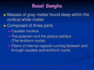
• The basal ganglia are four telencephalic nuclei (striatum = caudate, putamen, and ventral striatum):
o Caudate – located lateral to the lateral ventricle. It head is innervated by the prefrontal cortex, its body by the frontal and parietal cortex, and its tail by the occipital and temporal cortex.
o Putamen – separated from the caudate by the anterior limb of the internal capsule. It is innervated by M1 and caudal premotor areas (premotor and supplementary motor areas).
o Ventral striatum (includes nucleus accumbens) – innervated by limbic cortex (anterior cingulate and orbitofrontal) o Globuspallidus (internal GPi, and external GPe) – GPi is part of the direct pathway, GPe is part of the indirect pathway.
Striatum neurons are not very active and rarely fire quickly. GPiand GPe neurons are very active.
The two nuclei associated with the basal ganglia are the subthalamic nucleus (diencephalon, excites GPi), and substantianigra (mesencephalon, with a compact - SNpc - dopaminergic part and a reticulated - SNpr - GABAergic part). SNpr and GPi function in basically the same way, with GPi involved in body movement and SNpr involved in eye movements.
The primary motor area for the body is M1, for the eyes it is the Frontal Eye Fields (FEF). The basal ganglia handle these systems separately. Putamenhandles voluntary body movements, and the body of the caudate handles voluntary eye movements.
1 cortical motor neuron can be involved in many actions. To produce a particular movement, a population of neurons that each encodes a simple act must be activated with the appropriate strength, speed, etc. The basal ganglia function to activate such a specific program of movement.
• The basal ganglia are part of a circuit that loops from cortex → striatum → GPi or SNpr → thalamus → cortex. All areas of the cortex have some neurons that project to the striatum of the basal ganglia.
Different parts of the cortex project to different parts of the basal ganglia (ex: FEF → body of caudate, whereas M1/supplementary motor/premotor → putamen, and Limbic areas → Ventral Striatum). Other important areas include the Dorsolateral Prefrontal Cortex (executive function), and the Lateral Orbitofrontal and Anterior Cingulate Cortex (both involved with motivational drive).
• The axons from the cortex are excitatory, and they mostly innervate striatal medium spiny stellatecells. Most rostral cortical neurons project to the most rostralstriatal neurons.
The most caudal parts of the motor areas (spinal cord) directly control muscle actions, and more rostral parts become farther and farther removed (planning, motivation, decision making).
All outputs from the basal ganglia go through either: GPi, for body movements. SNprfor eye movements. Both of these regions have strong inhibitory (GABA) input to the ventral anterior (VA) nucleus or oral part of the ventral lateral (VLo) thalamus for body/eye movements.
For reflexive eye movements, the SNpr must also innervate the superior colliculus. For limbic functions, outputs project from the GPi to the mediodorsal (MD) thalamic nucleus.
• Cerebellar and basal ganglia are very similar, but they innervate different parts of the thalamus and cortex (cerebellum → M1 and caudal premotor….basal ganglia → rostralpremotor and prefrontal cortex).
Most striatal neurons are medium spiny neurons, long interneurons that take cortical input and project to the GPi or SNpr. A few are local interneurons using ACh.
Basal ganglia don’t initiate movement, but adjust it in its earliest stages. Most putamen neurons respond only after a movement has begun. GPineurons show early, broad activity, then later they become more focused (possibly from basal ganglia input?). This suggests that they adjust movements.
Half of basal ganglia neurons respond to the force/speed of movement, and an overlapping majority responds to direction. GPicells are also mostly direction-sensitive.
The direct pathway basically facilitates movement by liberating a motor program: Motor cortex → putamen/caudate ¬ GPi/SNr ¬ Thalmus → SMA/PMA/PFC(M1). The striatum relieves the GPi/SNr inhibition on the thalamus (disinhibition) to liberate motor programs.
Inhibition hyperpolarizes a neuron and makes more VG Na channels capable of being activated. So when the inhibition is relieved, the neuron is more likely to fire. Disrupting the direct pathway results in slow, weak movements (ex: bradykinesia in Parkinson’s).
• The indirect pathway inhibits thalamic output, ultimately suppressing competing motor programs: o Biggest effect: cortex → subthalamic nucleus → GPi/SNr ¬ thalamus (VA). This excites the GPi/SNr and increases its inhibition of the thalamus.
o Smaller effect: cortex → striatum ¬ GPe ¬ subthalamic nucleus → GPi/SNr ¬ thalamus (VA). Basically the striatum disinhibits the STN, allowing it to better activate the GPi/SNr and better inhibit the thalamus.
Disrupting the indirect pathway results in hyperkinetic symptoms (ex: proximal muscles contractions of Huntington’s). • Direct/indirect pathway input only activates or inhibits specific regions, not the whole thalamus. This enables you to activate and inhibit the necessary things to produce the desired response.
The compact part of the substantianigra (SNpc) release dopamine onto the caudate and putamen. D1 receptors increase the strength of glutamate receptors there, and D2 receptors reduce the strength of glutamate receptors.
o Direct pathway neurons preferentially express D1 receptors, so the direct pathway gets enhanced by DA and more movements are liberated. o Indirect pathway neurons preferentially express D2 receptors. so the indirect pathway gets weakened, resulting in less inhibition of suppressed motor programs.
To be able to activate a motor program when you need it, activation should be powerful and the suppression should be weak. DA accomplishes this. DA neurons are active when a reward for an action exceeds the expected reward, leading to increased activity in the active circuits.
DA neurons degenerate in Parkinson’s. It results in hyper- (tremor, heightened reflexes) and hypo-kinetic (bradykinesia) symptoms. The drug MPTP produces the same deficits.
Huntingon’s is characterized by weak voluntary and robust involuntary movements. It is autosomal dominant mutation in which the huntingtin protein has over 35 CAG repeats. This adds glutamines to the protein, destabilizing it and leading to apoptosis.
Early loss of medium spiny neurons in the striatum associated with the indirect pathway produce hyperkinetic activity (choreiform movements). Later loss of direct pathway neurons is associated with muscular rigidity.
This disease also shows anticipation, in which it shows earlier in subsequent generations. • Hemiballismus is a condition with sudden, dramatic involuntary movements of the limbs on one side. These are very forceful. It is produced by destruction of the contralateral STN.