Download
1 / 35
350 likes | 549 Views
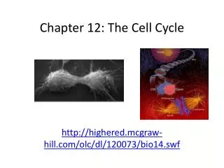
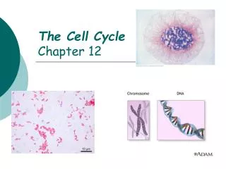
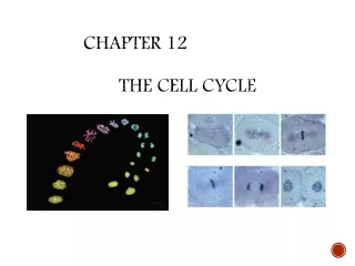
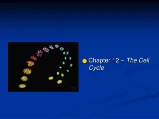
Chapter 12: The Cell Cycle. The Cell Cycle. Fig. 12-1. 100 m. (a) Reproduction. 200 m. Figure 12.2. Why do cells divide?. (b) Growth and development. 20 m. (c) Tissue renewal. Eukaryotes Have Chromosomes. Figure 12.3. 20 m.

E N D
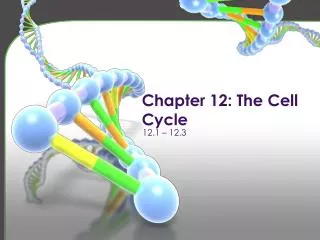
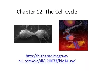
Chapter 12: The Cell Cycle
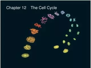
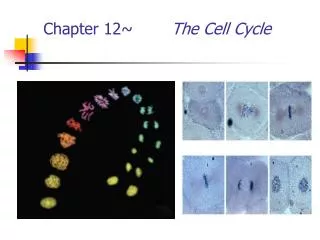
The Cell Cycle Fig. 12-1
100 m (a) Reproduction 200 m Figure 12.2 Why do cells divide? (b) Growth and development 20 m (c) Tissue renewal
Eukaryotes Have Chromosomes Figure 12.3 20 m
Chromosomes are duplicated before cell division! Figure 12.4 Sisterchromatids Centromere 0.5 m
ChromosomalDNA molecules Chromosomes Centromere 1 Chromosomearm Figure 12.5-1
ChromosomalDNA molecules Chromosomes Centromere 1 Chromosomearm Figure 12.5-2 Chromosome duplication(including DNA replication)and condensation 2 Sisterchromatids
ChromosomalDNA molecules Chromosomes Centromere 1 Chromosomearm Figure 12.5-3 Chromosome duplication(including DNA replication)and condensation 2 Sisterchromatids Separation of sisterchromatids intotwo chromosomes 3
Chromosome arrangement • Somatic cells – typical body cells • 46 chromosomes in 23 pairs (humans) • The chromosomes are not normally paired up • Each pair is called homologous chromosomes • Gametes – sex cells • 23 chromosomes (humans) • One member from each pair
Homologous chromosomes • 22 of the pairs (autosomes) are “true homologues” • One of each came from mom and dad • Identical in length and type of genes carried • Genes on each are slightly different • Sex chromosomes (23rd pair) don’t match up exactly (X vs Y)
5 m Pair of homologousduplicated chromosomes Figure 13.3b Centromere Sisterchromatids Metaphasechromosome
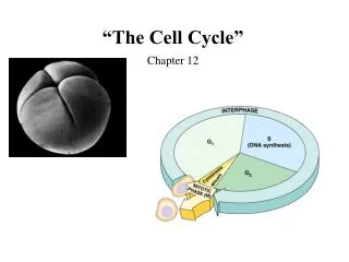
The Cell Cycle INTERPHASE S(DNA synthesis) G1 Figure 12.6 Cytokinesis G2 Mitosis MITOTIC(M) PHASE
Mitosis • Division of the nucleus • Results in two nuclei that have the same chromosome number as the parent • The two nuclei are genetically identical
Mitosis • Divided into 5 phases: • Prophase • Prometaphase • Metaphase • Anaphase • Telophase • Phases are determined by chromosome movement • Chromosome movement determined by mitotic spindle • Mitotic spindle controlled by centrosome
The Mitotic Spindle Centrosome Aster Metaphaseplate(imaginary) Sisterchromatids Microtubules Figure 12.8 Chromosomes Kineto-chores Centrosome 1 m Overlappingnonkinetochoremicrotubules Kinetochoremicrotubules 0.5 m
Prometaphase G2 of Interphase Prophase Fragments of nuclearenvelope Centrosomes(with centriole pairs) Early mitoticspindle Nonkinetochoremicrotubules Chromatin(duplicated) Aster Figure 12.7a Centromere Plasmamembrane Kinetochore Nucleolus Kinetochoremicrotubule Chromosome, consistingof two sister chromatids Nuclearenvelope
Metaphase Anaphase Telophase and Cytokinesis Nucleolusforming Metaphase plate Cleavagefurrow Figure 12.7b Nuclearenvelopeforming Spindle Centrosome atone spindle pole Daughterchromosomes
Figure 12.7c 10 m G2 of Interphase Prophase Prometaphase
Figure 12.7d 10 m Metaphase Anaphase Telophase and Cytokinesis
Mitosis in a Plant Cell Chromatincondensing Nucleus Figure 12.11 10 m Nucleolus Chromosomes Cell plate 3 4 5 2 Anaphase 1 Prophase Metaphase Telophase Prometaphase
Cytokenesis: Animal vs. Plant Cell (a) Cleavage of an animal cell (SEM) (b) Cell plate formation in a plant cell (TEM) Figure 12.10 100 m Vesiclesformingcell plate Cleavage furrow Wall of parent cell 1 m New cell wall Cell plate Daughter cells Contractile ring ofmicrofilaments Daughter cells
Figure 12.11 Binary Fission: Cell division in bacteria
Cell wall Origin ofreplication Plasma membrane E. coli cell Bacterial chromosome 1 Chromosomereplicationbegins. Two copies of origin Figure 12.12-1
Cell wall Origin ofreplication Plasma membrane E. coli cell Bacterial chromosome 1 Chromosomereplicationbegins. Two copies of origin Figure 12.12-2 2 Origin Origin Replicationcontinues.
Cell wall Origin ofreplication Plasma membrane E. coli cell Bacterial chromosome 1 Chromosomereplicationbegins. Two copies of origin Figure 12.12-3 2 Origin Origin Replicationcontinues. 3 Replicationfinishes.
Cell wall Origin ofreplication Plasma membrane E. coli cell Bacterial chromosome 1 Chromosomereplicationbegins. Two copies of origin Figure 12.12-4 2 Origin Origin Replicationcontinues. 3 Replicationfinishes. 4 Two daughtercells result.
Prokaryotic chromosomes LE 8-3b Colorized TEM 32,500
G1 checkpoint Figure 12.15 Controlsystem S G1 Cell Cycle Control G2 M M checkpoint G2 checkpoint
Checkpoints G0 G1 checkpoint Figure 12.16 G1 G1 (a) Cell receives a go-ahead signal. (b) Cell does not receive a go-ahead signal.
Cancer • Kills 1 out of 5 Americans • Disease of the cell cycle • Cells don’t respond to the normal cell cycle checkpoints • Cancer cells DO NOT exhibit density or anchorage dependence • Cancer cells divide out of control and can invade other parts of the body
Anchorage dependence Figure 12.19 Density-dependent inhibition Density-dependent inhibition 20 m 20 m (b) Cancer cells (a) Normal mammalian cells
Lymph vessel Figure 12.20 Tumor Bloodvessel Cancercell Glandulartissue Metastatictumor A tumor growsfrom a singlecancer cell. Cancer cells invade neighboringtissue. Cancer cells spreadthrough lymph andblood vessels to other parts of the body. Cancer cells may survive and establisha new tumor in another part of the body. 1 2 3 4
P HA R E S T E N I G1 S Cytokinesis Mitosis G2 Figure 12.UN01 MITOTIC (M) PHASE Prophase Telophase andCytokinesis Prometaphase Anaphase Metaphase