Download
1 / 50
520 likes | 605 Views
Chapter 12: The Cell Cycle. 12.1 – 12.3. Overview. Living things are known to be living things because of the characteristics of life , reproduction being one of them. There are two kinds of reproduction: sexual and asexual (cell division).

E N D
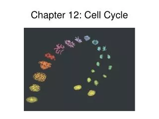
Chapter 12: The Cell Cycle 12.1 – 12.3
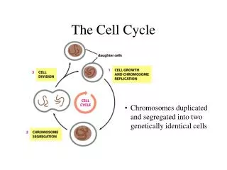
Overview • Living things are known to be living things because of the characteristics of life, reproduction being one of them. • There are two kinds of reproduction: sexual and asexual (cell division). • Cell division functions in reproduction, growth, and repair. • When a unicellular organism reproduces the end result is two separate organisms. • Cell division on a large scale can produce progeny for some multicellular organisms. • Eg. cuttings for plants • Cell division also enables a multicellular organism to develop from a single fertilized egg or zygote. • Cell division functions to repair and renew cells that die from normal wear and tear or accidents. • This is part of the cell cycle, the life of a cell from its origin in the division of a parent cell until its own division into two.
12.1 Cell Division Results in Genetically Identical Daughter Cells
Genetic Material • Cell division requires the distribution of identical genetic material – DNA – to two daughter cells. • Remarkably DNA can be passed along from one generation to the next with no dilution. • A dividing cell duplicates its DNA, allocates the two copies to opposite sides of the cell, and splits down the middle into two daughter cells. • The genome of a cell is the complete genetic information – prokaryotes usually have a single long DNA molecule, eukaryotes usually have several DNA molecules. • In humans, dividing cells must copy 2 m of DNA and separate the two copies so that each daughter cell ends up with a complete genome. • DNA molecules are packaged into chromosomes. • Every eukaryotic species has a characteristic number of chromosomes in each cell nucleus. • Human somatic cells (body cells) have 46 chromosomes, 23 from each parent. • Human gametes (sperm or eggs) have 23 chromosomes.
Chromosomes • Eukaryotic chromosomes are made of chromatin, a complex of DNA and associated proteins. • Each single chromosome contains one long, linear DNA molecule carrying hundreds or thousands of genes, the units that specify an organism’s inherited traits. • The associated proteins maintain the structure of the chromosome and help control gene activity. • When a cell is not dividing, each chromosome is in the form of a long, thin chromatin fiber. • Each duplicated chromosomes consists of two sister chromatids, which contain identical copies of the chromosome’s DNA. • The chromatids are initially attached by adhesive proteins along their lengths. • As the chromosomes condense, the region where the chromatids connect shrinks to a narrow area, the centromere. • Later in cell division, the sister chromatids are pulled apart and repackaged into two new nuclei at opposite ends of the parent cell. • Once the sister chromatids separate, they are considered individual chromosomes.
Mitosis • Mitosis: the formation of the two daughter nuclei, is usually followed by the division of the cytoplasm, cytokinesis. • These processes start with one cell and produce two cells that are genetically identical to the original parent cell. • Each of us inherited 23 chromosomes from each parent: one set in an egg, one set in a sperm. • The fertilized egg (zygote) undergoes cycles of mitosis and cytokinesis to produce a fully developed multicellular human made up of 200 trillion somatic cells. • This continues all day every day to replace dead and damaged cells. • Essentially, these processed produce clones – cells with identical genetic information.
Meiosis • In contrast to mitosis, gametes (egg and sperm) are produced only in gonads (ovaries or testes) by a variation of cell division called meiosis. • Meiosis yields four nonidentical daughter cells, each with half the chromosomes of the parent. • In humans, meiosis reduces the number of chromosomes from 46 to 23. • Fertilization fuses two gametes together and doubles the number of chromosomes to 46 again.
12.2 The Mitotic Phase Alternates with Interphase in the Cell Cycle
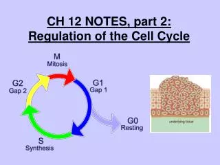
Phases • The mitotic phase (M) of the cell cycle alternates with the much longer interphase. • The M phase includes mitosis and cytokinesis. • Interphase accounts for 90% of the cell cycle. • During interphase, the cell grows by producing proteins and cytoplasmic organelles, copies it chromosomes, and prepares for cell division. • Interphase and three subdivisions: the G1 phase (“first gap”), the S phase (“synthesis”), and the G2 phase (“second gap”). • During all three subphases, the cell grows by producing proteins and cytoplasmic organelles such as mitochondria and ER. • Chromosomes are duplicated ONLY during the S phase.
Time Frames • The daughter cell may repeat the cycle. • A typical human cell might divide once every 24 hours. • Of this time, the M phase would last less than an hour, while the S phase might take 10-12 hours, or half the cycle. • The rest of the time would be divided between the G1 and G2 phases. • The G1 phase varies most in length from cell to cell.
Phases of Mitosis • Mitosis is broken into 5 subphases: prophase, prometaphase, metaphase, anaphase, and telophase. • In late interphase, the chromosomes have been duplicated but are not condensed. • A nuclear membrane binds to the nucleus, which contains one or more nucleoli. • The centrosome has replicated to form two centrosomes. • In animal cells, each centrosome features two centrioles.
Prophase • In prophase, the chromosomes are tightly coiled, with sister chromatids joined together. • The nuclei disappear and the mitotic spindle begins to form. • The mitotic spindle is composed of centrosomes and the microtubules that extend from them. • The radial arrays of shorter microtubules that extend from the centrosomes are called asters. • The centrosomes move away from each other, pulled by lengthening microtubules.
Prometaphase • During prometaphase, the nuclear envelope fragments and microtubules from the spindle interact with the condensed chromosomes. • Each of the two chromatids of a chromosome has a kinetochore, a specialized protein structure located at the centromere. • Kinetochore microtubules from each pole attach to one of two kinetochores. • Nonkinetochore microtubules interact with those from opposite ends of the spindle. • The spindle fibers push the sister chromatids until they are all arranged at the metaphase plate, an imaginary plane equidistant from the poles that defines metaphase.
Anaphase and Telophase • In anaphase, the centromeres divide, separating the sister chromatids. • Each is now pulled toward the pole where the spindle fiber is attached. • By the end, the two poles have equidistant collections of chromosomes. • In telophase, daughter nuclei begin to form at the two poles. • Nuclear envelopes arise from the fragments of the parent cell’s nuclear envelope and other portions of the endomembrane system. • The chromosomes become less tightly coiled.
Cytokinesis • Cytokinesis is division of the cytoplasm. • Also known as cleavage, the process is usually underway by the end of telophase. • In animal cells, cytokinesis involves the formation of a cleavage furrow, which pinches the cell in two. • In plant cells, vesicles derived from the Golgi apparatus produce a cell plate at the middle of the cell.
Interphase: A Closer Look • The mitotic spindle (microtubules and associated proteins) is a major driving force in mitosis. • As the spindle assembles during prophase, the elements come from a partial disassembly of the cytoskeleton. • The spindle fibers elongate by incorporating more subunits of the protein tubulin. • Assembly of the spindle microtubules starts in the centrosome. • The centrosome (microtubule-organized center) is a nonmembranous organelle that organize the cell’s microtubules. • In animal cells, the centrosome has a pair of centrioles at the center, but the centrioles are not essential for cell division. • During interphase, the single centrosome replicates to form two centrosomes. • As mitosis starts, the two centrosomes are located near the nucleus. • As the spindle microtubules grow from them, the centrioles are pushed apart. • By the end of prometaphase, they are at opposite ends of the cell.
Mitotic Cycle: A Closer Look • An aster, (radial array of short microtubules) extends from each centrosome. • The spindle includes the centrosomes, the spindle microtubules, and the asters. • Each sister chromatid has a kinetochore of proteins and chromosomal DNA at the centromere. • The kinetochores of the joined sister chromatids face in opposite directions. • During prometaphase, some spindle microtubules (kinetochore microtubules) attach to the kinetochores. • When a chromosome’s kinetochore is “captured” by microtubules, the chromosome moves toward the pole the microtubules came from. • When microtubules attach to the other pole, this movement stops and a tug-of-war ensues. • Eventually, the chromosome settles midway between the two poles of the cell on the metaphase plate. • Nonkinetochore microtubules from opposite poles overlap and interact with each other. • By metaphase the microtubules of the asters have grown and are in contact with the plasma membrane. • The spindle is now complete. • Anaphase commences when the protein holding the sister chromatids together are inactivated. • One the chromosomes are separated, they move towards the opposite poles of the cell.
Kinetochores • It’s not quite clear how kinetochores function in the movement of the chromosomes. • One hypothesis is that chromosomes are “reeled in” by the shortening of microtubules at the spindle pores. • Experimental evidence supports the hypothesis that motor proteins (called dynein) on the kinetochore “walk” the attached chromosome along the microtubule toward the nearest pole. • Meanwhile, the excess microtubule sections depolymerize at their kinetochore ends. • Function: • Lengthening the cell along the axis defined by the poles. • The microtubules interdigitate and overlap across the metaphase plate. • During anaphase, the are of overlap is reduced as dynein walks them away, using ATP. • The microtubules lengthen by the addition of new tubulin monomers to their overlapping ends, allowing continued overlap.
Cytokinesis: A Closer Look • Cytokinesis follows mitosis. • In animal cells, it is also called cleavage. • The first sign of cleavage is the appearance of a cleavage furrow in the cell surface near the old metaphase plate. • On the cytoplasmic side of the cleavage furrow is a contractile ring of actin microfilaments associated with molecules of the motor protein myosin. • Contraction of the ring pinches the cell in two. • Cytokinesis in plant is completely different. • During telophse, vesicles from the Golgi coalesce at the metaphase plate, forming a cell plate. • The plate enlarges until its membranes fuse with the plasma membrane at the perimeter. • The contents of the vesicles form new cell wall material between the daughter cells.
Binary Fission Mitosis • Prokaryotes reproduce by binary fission, not mitosis. • Most bacterial genes are located on a single bacterial chromosome that consists of a circular DNA molecule and associated proteins (a plasmid). • While bacteria are smaller and simpler than eukaryotic cells, they still have large amounts of DNA that must be copied and distributed equally to two daughter cells. • The circular bacterial chromosome is highly folded and coiled in the cell. • In binary fission, chromosome replication begins at one point in the circular chromosome, the origin of replication site, producing two origins. • As the chromosome continues to replicate, one origin moves towards each end of the cell. • While the chromosome is replicating, the cell elongates. • When replication is complete, its plasma membrane grows inward to divide the parent cell into two daughter cells, each with a complete genome.
Mechanism of Binary Fission • Researchers have developed methods to allow them to observe the movement of bacterial chromosomes. • It’s similar to the poleward movement of the centromere regions of eukaryotic chromosomes. • However, bacterial chromosomes lack visible mitotic spindles or even microtubules. • The mechanism is still unclear, but several proteins have been identified and play important roles. • Mitosis had its origin in bacterial binary fission. • Some of the proteins involved in binary fission are related to eukaryotic proteins. • Two are related to eukaryotic tubulin and actin proteins. • Possible intermediate evolutionary steps are seen in the division of two types of unicellular algae. • In dinoflagellates, replicated chromosomes are attached to the nuclear envelope. • In diatoms, the spindle develops within the nucleus. • In most eukaryotic cells, the nuclear envelope breaks down and a spindle separates the chromosomes.
12.3 The Cell Cycle is Regulated by a Molecular Control System
Timing • The timing and rates of cell division in different parts of an animal or plant are crucial for normal growth, development, and maintenance. • The frequency caries with cell type: • Some human cells divide frequently throughout life (skin cells). • Other have the ability to divide, but keep it in reserve (liver cells). • Mature nerve and muscle cells do not appear to divide at all after maturity. • Investigation of the molecules mechanisms regulating these differences provide important insights into the operation of normal cells, and may also explain how cancer cells escape controls.
Cytoplasmic Signals • Some of the initial evidence for the hypothesis that specific chemical signals in the cytoplasm came from experiments in which cultured mammalian cells at different phases of the cell cycle were fused to form a single cell with two nuclei. • Fusion of an S phase cell and a G1 phase cell induces the G1 nucleus to start S phase. • This suggests that chemicals present in the S phase nucleus stimulated the fused cell. • Fusion of a cell in mitosis (M phase) with one in interphase (even G1 phase) induces the second cell to enter mitosis. • The sequential events of the cell cycle are directed by a distinct cell cycle control system. • Cyclically operating molecules trigger and coordinate key events in the cell cycle. • The control cycle has a built-in clock, but it is also regulated by external adjustments and internal controls.
Checkpoints • A checkpoint in the cell cycle is a critical point where stop and go-ahead signals regulate the cycle. • The signals are transmitted within the cell by signal transduction pathways. • Animal cells generally have built-in stop signals that halt the cell cycle at checkpoints until overridden by go-ahead signals. • Many signals registered at checkpoints come from cellular surveillance mechanisms. • These indicate whether key cellular processes have been completed correctly. • Checkpoints also register signals from outside the cell. • Three major checkpoints are found in the G1, G2, and M phases.
Most Important Checkpoint • For most cells in mammals and other animals, the G1 checkpoint, the “restriction point”, is the most important. • If the cell receives a go-ahead signal at the G1 checkpoint, it usually completes the cell cycle and divides. • If it does not receive a go-ahead signal, the cell exits the cycle and switches to a nondividing state, the G0 phase. • Most cells in the human body are in this phase. • Liver cells can be “called back” to the cell cycle by external cues, such as growth factors and released during injury. • Highly specialized nerve and muscle cells never divide. • What does this mean for these cells in the long run?
Fluctuations • Rhythmic fluctuations in the abundance and activity of cell cycle control molecules pace the events of the cell cycle. • These regulatory molecules include protein kinases that activate or deactivate other proteins by phosphorylating them. • These kinases are present in constant amounts but require attachment of a second protein, a cyclin, to become activated. • Levels of cyclin proteins fluctuate cyclically. • Because of the requirement for binding of a cyclin, the kinases are called cyclin-dependent kinases, or Cdks. • Cyclin level rise sharply throughout interphase, and then fall abruptly during mitosis.
Corresponding Peaks • Peaks in the activity of one cyclin-Cdk complex, MPF, correspond to peaks in cyclin concentration. • MPF (“maturation-promoting factor” or “M phase promoting factor”) triggers the cell’s passage past the G2 checkpoint to the M phase. • MPF promotes mitosis by phosphorylating a variety of other protein kinases. • MPF stimulates fragmentation of the nuclear envelope by phosphorylation of various proteins of the nuclear lamina. • It also triggers the breakdown of cyclin, dropping cyclin and MPF levels during mitosis and inactivation MPF. • The noncyclin part of MPF, the Cdk, persists in the cell in inactive form until it associates with new cyclin molecules synthesized during the S and G2 phases of the next round of the cycle. • At least three Cdk proteins and several cyclins regulate the key G1 checkpoint. • Similar mechanisms are also involved in driving the cell cycle past the M phase checkpoint
Internal and External Cues • While research scientists know that active Cdks function by phosphorylation proteins, the identity of all these proteins is still under investigation. • Scientists do not yet know what Cdks actually do in most cases. • Some steps in the signaling pathways that regulate the cell cycle are clear. • Some signals originate inside the cell, others outside.
M Phase Checkpoint • All chromosomes have to be properly attached to the spindle at the metaphase plate before anaphase in order to pass the M phase checkpoint. • This ensures that daughter cell do not end up with missing or extra chromosomes (nondisjunction). • A signal to delay anaphase originates at kinetochores that have not yet attached to spindle microtubules. • This keeps the anaphase-promoting complex (APC) in an inactive state. • When all kinetochores are attached the APC activates, triggering breakdown of cyclin and inactivation of proteins holding sister chromatids together. • A variety of external chemical and physical factors can influences cell division. • Eg. Cells fail to divide if an essential nutrient is left out of the culture medium.
PDGF • Particularly important for mammalian cells are growth factors, proteins released by one group of cells that stimulate other cells to divide. • Eg. Platelet-derived growth factors (PDGF) produced by platelet blood cells bind to tyrosine-kinase receptors of fibroblasts (connective tissue cells). • This triggers a signal-transduction pathway that allows cells to pass the G1 checkpoint and divide. • Each cell type probably responds specifically to a certain growth factor or combination of factors. • The role of PDGF is easily seen in cell culture. • Fibroblasts in culture will only divide in the presence of a medium that also contains PDGF. • In a living organism, PDGF is released in the vicinity of an injury. • The resulting proliferation of fibroblasts helps heal the wound.
External Physical Factors • At least 50 different growth factors can trigger specific cells to divide. • The effect of an external physical factor on cell division can be seen in density-dependent inhibition of cell division. • Cultured cells normally divide until they form a single layer on the inner surface of the culture container. • If a gap is created, the cells will grow to fill the gap. • At high densities, the amount of growth factors and nutrients is insufficient to allow continued cell growth. • Most animal cells also exhibit anchorage dependence for cell division. • To divide, they must be anchored to a substratum, typically the extracellular matrix of a tissue. • Control appears to be mediated by pathways involving plasma membrane proteins and elements of the cytoskeleton linked to them. • Cancer cells exhibit neither density-dependent inhibition nor anchorage dependence.
Cancer Cells • Cancer cells divide excessively and invade other tissues because they are free of the body’s control mechanisms. • Cancer cells do not stop dividing when growth factors are depleted. • This is either because a cancer cell manufactures its own growth factors, has an abnormality in the signaling pathway, or has an abnormal cell cycle control system. • In and when cancer cells stop dividing, they do so at random points, not at when the normal checkpoints in the cell cycle. • Cancer cells may divide indefinitely if they have a continual supply of nutrients. • In contrast, nearly all mammalian cells divide 20-50 times under culture conditions before they stop, age, and die. • Cancer cells may be “immortal”. • HeLa cells from a tumor removed from a woman (Henrietta Lacks) in 1951 are still reproducing in culture.
Abnormal Behavior • The abnormal behavior of cancer cells begins when a single cells in a tissue undergoes a transformation that converts it from a normal cell to a cancer cell. • Normally, the immune system recognizes and destroys transformed cells. • However, cells that evade destruction proliferate to form a tumor, a mass of abnormal cells. • If the abnormal cells remain at the originating site, the lump is called a benign tumor. • Most do not cause serious problems and can be fully removed by surgery. • In a malignant tumor, the cells become invasive enough to impair the functions of one or more organs.
It All Starts Falling Apart • In addition to chromosomal and metabolic abnormalities, cancer cells often lose attachment to nearby cells, are carried by the blood and lymph system to other tissues, and start more tumors in an event called metastasis. • Cancer cells have an unusual number of chromosomes, their metabolism may be disabled, and they may cease to function in any constructive way. • Cancer cells may secrete signal molecules that cause blood vessels to grow toward the tumor. • Treatments for metastasizing cancers include high-energy radiation and chemotherapy with toxic drugs. • These treatments target actively dividing cells. • Chemotherapeutic drugs interfere with specific steps in the cells cycle. • Eg. Taxol prevents mitotic depolymerization, preventing cells from proceeding past metaphase. • The side effects of chemotherapy are due to the drug’s effects on normal cells. • Researchers are beginning to understand how a normal cell is transformed into a cancer cell. • The causes are diverse, but cellular transformation always involves the alteration of genes that influence the cell cycle control system.
Nerd Alert • http://safeshare.tv/w/BSkSJfOHIk • http://safeshare.tv/w/rxzhjlxYuF • http://safeshare.tv/w/eCxemeWgPQ • Henrietta Lacks: http://www.bbc.co.uk/blogs/adamcurtis/2010/06/the_undead_henrietta_lacks_and.html