Download
1 / 50
610 likes | 929 Views
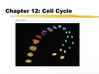
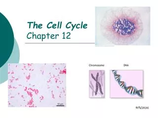
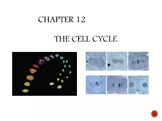
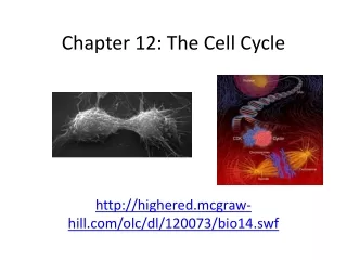
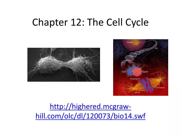
Chapter 12: The Cell Cycle. http://highered.mcgraw-hill.com/olc/dl/120073/bio14.swf. Omnis cellula e cellula From every cell a cell – Rudolf Virchow. Cell division : reproduction of cells

E N D
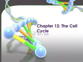
Chapter 12: The Cell Cycle http://highered.mcgraw-hill.com/olc/dl/120073/bio14.swf
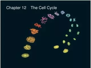
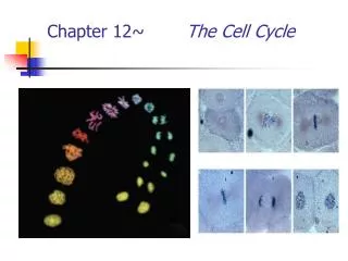
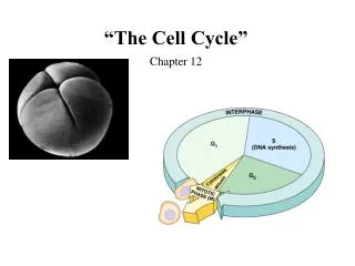
Omnis cellula e cellulaFrom every cell a cell – Rudolf Virchow • Cell division: reproduction of cells • Cell cycle: life of a cell from the time it is first formed from a dividing parent cell until it divides into 2 daughter cells • Mitosis: nuclear division within a cell, followed by cytokinesis • Cytokinesis: division of the cytoplasm
Getting the right stuff • What is passed on to daughter cells? • exact copy of genetic material = DNA • mitosis • organelles, cytoplasm, cell membrane, enzymes • cytokinesis chromosomes (stained orange)in kangaroo rat epithelial cell notice cytoskeleton fibers
Mitosis amoeba • Mitosis functions in • Reproduction of single-celled organisms • Growth • Repair • Regeneration • In contrast, meiosis produces gametes in sexually-reproducing organisms • Contains half the number of chromosomes of a somatic cell
M anaphase metaphase telophase prophase C G2 interphase (G1, S, G2 phases) mitosis (M) cytokinesis (C) G1 S Frequency of cell division • Frequency of cell division varies by cell type • embryo • cell cycle < 20 minute • skin cells • divide frequently throughout life • 12-24 hours cycle • liver cells • retain ability to divide, but keep it in reserve • divide once every year or two • mature nerve cells & muscle cells • do not divide at all after maturity • permanently in G0
Organization of the Genetic Material • Cell division results in genetically identical daughter cells • This requires DNA replication followed by division of the nucleus • Genome: genetic content of the cell • Prokaryotic cells have circular DNA Circular DNA: a single long molecule • Eukaryotic cells contain a number of DNA molecules specific to different species One eukaryotic cell has about 2 meters of DNA
ACTGGTCAGGCAATGTC Organizing DNA DNA • DNA is organized in chromosomes • double helix DNA molecule • wrapped around histone proteins • like thread on spools • DNA-protein complex =chromatin • organized into long thin fiber • condensed further during mitosis histones chromatin duplicated mitotic chromosome
Distribution of Chromosomes During Cell Division • Non-dividing cells contain chromatin • In human cells there are 46 strands of chromatin, or 23 corresponding pairs of strands • Dividing cells contain chromosomes • Chromosomes becomes condensed prior to mitosis • Chromosomes contain sister chromatids bound by a narrow centromere • Kinetochore: a structure of proteins associated with specific sections of chromosomal DNA at the centromere
0.5 µm A eukaryotic cell has multiplechromosomes, one of which is represented here. Before duplication, each chromosomehas a single DNA molecule. Chromosomeduplication(including DNA synthesis) Once duplicated, a chromosomeconsists of two sister chromatidsconnected at the centromere. Eachchromatid contains a copy of the DNA molecule. Centromere Sisterchromatids Separation of sister chromatids Mechanical processes separate the sister chromatids into two chromosomes and distribute them to two daughter cells. Centromeres Sister chromatids Figure 12.4 Chromosome Structure
Mitotic Phase Alternates with Interphase in Cell Cycle • Mitosis: M Phase includes mitosis and cytokinesis • Prophase • Prometaphase • Metaphase • Anaphase • Telophase • Interphase: 90% of cell cycle; cell grows, producing organelles and proteins • G1: First gap • S: Synthesis, chromosome replication • G2: Second gap; completes preparation for cell division I’m working here! Time to Divide
INTERPHASE S(DNA synthesis) G1 CytokinesisMitosis G2 MITOTIC(M) PHASE In 24 hours… • Mitosis: 1 hour • G1: 5-6 hours (most variable) • S: 10-12 hours • G2: 4-6 hours
Aster Centrosome MetaphasePlate Sisterchromatids Kinetochores Overlappingnonkinetochoremicrotubules Kinetochores microtubules 0.5 µm Microtubules Chromosomes Centrosome 1 µm Mitotic Spindle is Necessary for Nuclear Division • Mitotic spindle: used to segregate sister chromatids in anaphase • Consists of microtubules and proteins • Microtubules are able to change in length • Elongate by adding Tubulin subunits • Shorten by loss of Tubulin subunits
Mitotic Spindle • Interphase:the centrosome is replicated and the two centrosomes remain paired near the nucleus • Centrosome or Microtubule Organizing Center (MTOC): contains two centrioles • Prophase: spindle formation • Early Prometaphase: migration of centrosomes to poles, spindle microtubules grow • Late Prometaphase: centrosomes are at the poles, asters form • Aster: radial array of short microtubules • The mitotic spindle includes the centrosomes, spindle microtubules and asters
Cellular Changes Indicate M- Phase Initiation G2 of Interphase: nuclear envelope intact • Nucleus and nucleolus present • Centrosome Replication • Animal cells contain centrioles • Chromosomes are not condensed and therefore not individually visible
Prophase • Nucleoli disappear • Chromosomes condense • Sister chromatids form • Two genetically identical arms joined by the centromere and cohesin proteins • Mitotic spindle forms using microtubules; asters form • Centrosome migration as microtubules lengthen
Prometaphase • Nuclear envelope fragments and microtubules invade nuclear area • Nonkinetochore microtubules elongate • Chromosomes condense further and gain kinetochore proteins • Spindle fibers (microtubules) interact with kinetochores
Mitotic Spindle Prometaphase: • Kinetochore: protein structure assiciated with specific sections of the chromosomal DNA at the centromere • Kinetochore microtubules: attach kinetochore to spindle • Metaphase Plate: created as microtubules attach to kinetochore, creation indicates Metaphase start
Metaphase • Longest stage of mitosis • Can last up to 20 minutes • Metaphase plate: site of chormosome alignment • Centrosomes are at poles • Sister chromatidkinetochores attach to kinetochore microtubules
Anaphase • Shortest stage of mitosis • Cohesins between sister chromatids are cleaved by enzymes • Sister chromatids are now separate chromosomes • Spindle fibers shorten causing chromosomes to move to opposite poles • Spindles shorten (tubulin breakdown) at kinetochoreends • Nonkinetochoremicrotubules elongate cell
Chromosome movement • Kinetochores use motor proteins that “walk” chromosome along attached microtubule • microtubule shortens by dismantling at kinetochore (chromosome) end
Telophase • Daughter nuclei form in cell • Chromosomes loosen and become less dense • Nucleoli reappear
Cytokinesis • Occurs in animal cells only • Relies on cleavage marked by a cleavagefurrow • Cytoplasmic side of cleavage furrow contains actin microfilaments and myosin • As the actin and myosin interact the ring contracts and the cleavage furrow deepens
Cellular Division in Plant Cells • Plant cells form a cell plate following mitosis • Golgi vesicles contain cell wall materials and migrate toward the center of the cell – forming the cell plate, a new cell wall
Binary Fission • Asexual eukaryotes can utilize mitosis for reproductive purposes – this is called binary fission • Asexual prokaryotes perform binary fission that does not involve mitosis
Evolution of Mitosis Attach to nuclear envelope • Some proteins were highly conserved • In prokaryotes, protein resembling eukaryotic actin may help with cell division • Tubulin-Like proteins may help separate daughter cells Reinforce spatial arrangement of nucleus Spindle inside nucleus
Cell Cycle Regulation • Cytoplasmicregulators as shown in Mammalian cell Fusion experiment by Johnson and Rao (1970)
Cell Cycle Control System • Control systems vary cell to cell • Skin cells divide frequently, liver cells only divide when needed (after injury), and nerve and muscle cells never divide • Cytoplasmic molecules signal cell cycle as shown by Johnson and Rao • Cell cycle events are regulated cyclicly • These are referred to as Cell Cycle Checkpoints
Cell Cycle Control Systems • Cell Cycle Checkpoint: control point in cell cycle where stop and go-ahead signals can regulate the cycle; relies on signal transduction pathways controlled by internal and external molecular signals • G1 checkpoint: acts as a mammalian restriction point • “Go Signal” permits G1, S, G2 and M • “Stop signal” causes G0 phase: non-dividing state • G2 checkpoint • M checkpoint
Checkpoint control system • 3 major checkpoints: • G1/S • can DNA synthesis begin? • G2/M • has DNA synthesis been completed correctly? • commitment to mitosis • spindle checkpoint • are all chromosomes attached to spindle? • can sister chromatids separate correctly?
G0 phase • G0 phase • non-dividing, differentiated state • most human cells in G0 phase • liver cells • in G0, but can be “called back” to cell cycle by external cues • nerve & muscle cells • highly specialized • arrested in G0 & can never divide
Cell Cycle Clock • The rate of cell cycle is controlled by two proteins: • Cyclins • Cyclin-dependent kinases (Cdks)
Cell Cycle Clock • Kinases: activate or inactivate other proteins via phosphorylation • Inactive most of the time • Kinases are activated by cyclins • Cyclins: regulatory proteins that have a cyclic (fluctuating) concentration in the cell • Cyclin-dependent kinases(Cdks) • Activity fluctuates with cyclin concentration
Cell Cycle Clock • MPF: maturation-promoting factor or M-phase promoting factor • MPF is a Cyclin-Cdk complex • Triggers cell passage from G2 to M • G2 checkpoint: As cyclin builds during G2 it binds with Cdk • Resulting MPF phosphorylates proteins, initiating Mitosis
During anaphase, MPF inhibits itself by destroying its own cyclin • Cdk persists in the cell
Stop and Go Signals • Internal signals • M Phase checkpoint relies on kinetochore signaling • Allows for enzymatic cleavage of cohesins • External factors • Nutrients • Growth Factor Dependency • Density-dependent inhibition • Anchorage dependence
External signals • Growth factors • coordination between cells • protein signals released by body cells that stimulate other cells to divide • density-dependent inhibition • crowded cells stop dividing • each cell binds a bit of growth factor • not enough activator left to trigger division in any one cell • anchorage dependence • to divide cells must be attached to a substrate • “touch sensor” receptors
Growth factor signals growth factor nuclear pore nuclear membrane P P cell division cell surface receptor Cdk E2F protein kinase cascade P chromosome P Rb P E2F Rb nucleus cytoplasm
Example of a Growth Factor • Platelet Derived Growth Factor (PDGF) • made by platelets in blood clots • binding of PDGF to cell receptors stimulates cell division in connective tissue • heal wounds
Growth Factors and Cancer • Growth factors can create cancers • proto-oncogenes • normally activates cell division • growth factor genes • become oncogenes (cancer-causing) when mutated • if switched “ON” can cause cancer • example: RAS (activates cyclins) • tumor-suppressor genes • normally inhibits cell division • if switched “OFF” can cause cancer • example: p53
Cancer & Cell Growth • Cancer is essentially a failure of cell division control • unrestrained, uncontrolled cell growth • What control is lost? • lose checkpoint stops • gene p53 plays a key role in G1/S restriction point • p53 protein halts cell division if it detects damaged DNA • options: • stimulates repair enzymes to fix DNA • forces cell into G0 resting stage • keeps cell in G1 arrest • causes apoptosis of damaged cell • ALL cancers have to shut down p53 activity p53 is theCell CycleEnforcer p53 discovered at Stony Brook by Dr. Arnold Levine
p53 — master regulator gene NORMAL p53 p53 allows cells with repaired DNA to divide. p53 protein DNA repair enzyme p53 protein Step 2 Step 1 Step 3 DNA damage is caused by heat, radiation, or chemicals. Cell division stops, and p53 triggers enzymes to repair damaged region. p53 triggers the destruction of cells damaged beyond repair. ABNORMAL p53 abnormal p53 protein cancer cell Step 2 Step 1 Step 3 The p53 protein fails to stop cell division and repair DNA. Cell divides without repair to damaged DNA. DNA damage is caused by heat, radiation, or chemicals. Damaged cells continue to divide. If other damage accumulates, the cell can turn cancerous.
Development of Cancer • Cancer develops only after a cell experiences ~6 key mutations (“hits”) • unlimited growth • turn on growth promoter genes • ignore checkpoints • turn off tumor suppressor genes (p53) • escape apoptosis • turn off suicide genes • immortality = unlimited divisions • turn on chromosome maintenance genes • promotes blood vessel growth • turn on blood vessel growth genes • overcome anchor & density dependence • turn off touch-sensor gene It’s like anout-of-controlcar with manysystems failing!
What causes these “hits”? • Mutations in cells can be triggered by • UV radiation • chemical exposure • radiation exposure • heat • cigarette smoke • pollution • age • genetics
Tumors • Mass of abnormal cells • Benign tumor • abnormal cells remain at original site as a lump • p53 has halted cell divisions • most do not cause serious problems &can be removed by surgery • Malignant tumor • cells leave original site • lose attachment to nearby cells • carried by blood & lymph system to other tissues • start more tumors =metastasis • impair functions of organs throughout body
Cancer Cells • Benign tumors: can be removed by surgery • Malignant tumors: invade surrounding tissues and organs, and often metastasize • Metastasis: spread of cancer to far locations in the body • Treatment options • Radiation: destroys cancer cells • Chemotherapy: medication that targets rapidly dividing cells (including cancer cells) • Ex: Taxol – stops dividing cells by prevents microtubule depolymerization in metaphase, but also affects cells that naturally divide often such as intestinal cells and skin cells of hair follicles
Traditional treatments for cancers • Treatments target rapidly dividing cells • high-energy radiation • kills rapidly dividing cells • chemotherapy • stop DNA replication • stop mitosis & cytokinesis • stop blood vessel growth