Download
1 / 35
350 likes | 550 Views
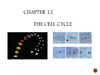
Chapter 12 The Cell Cycle. 2. Fig. 12-UN1. INTERPHASE. G 1. S. Cytokinesis. Mitosis. G 2. MITOTIC (M) PHASE. Prophase. Telophase and Cytokinesis. Prometaphase. Anaphase. Metaphase. Fig. 12-8. EXPERIMENT. Kinetochore. Spindle pole. Mark. RESULTS. CONCLUSION. Chromosome

E N D
Fig. 12-UN1 INTERPHASE G1 S Cytokinesis Mitosis G2 MITOTIC (M) PHASE Prophase Telophase and Cytokinesis Prometaphase Anaphase Metaphase
Fig. 12-8 EXPERIMENT Kinetochore Spindle pole Mark RESULTS CONCLUSION Chromosome movement Kinetochore Tubulin subunits Motor protein Microtubule Chromosome
Binary Fission • Prokaryotes (bacteria and archaea) reproduce by a type of cell division called binary fission • In binary fission, the chromosome replicates (beginning at the origin of replication), and the two daughter chromosomes actively move apart
Fig. 12-11-4 Cell wall Origin of replication Plasma membrane E. coli cell Bacterial chromosome Two copies of origin Origin Origin
The Evolution of Mitosis • Since prokaryotes evolved before eukaryotes, mitosis probably evolved from binary fission • Certain protists exhibit types of cell division that seem intermediate between binary fission and mitosis
Fig. 12-12 Bacterial chromosome (a) Bacteria Chromosomes Microtubules Intact nuclear envelope (b) Dinoflagellates Kinetochore microtubule Intact nuclear envelope (c) Diatoms and yeasts Kinetochore microtubule Fragments of nuclear envelope (d) Most eukaryotes
Concept 12.3: The eukaryotic cell cycle is regulated by a molecular control system • The frequency of cell division varies with the type of cell • These cell cycle differences result from regulation at the molecular level
Evidence for Cytoplasmic Signals • The cell cycle appears to be driven by specific chemical signals present in the cytoplasm • Some evidence for this hypothesis comes from experiments in which cultured mammalian cells at different phases of the cell cycle were fused to form a single cell with two nuclei
EXPERIMENT Experiment 1 Experiment 2 G1 S G1 M RESULTS M S S M When a cell in the S phase was fused with a cell in G1, the G1 nucleus immediately entered the S phase—DNA was synthesized. When a cell in the M phase was fused with a cell in G1, the G1 nucleus immediately began mitosis—a spindle formed and chromatin condensed, even though the chromosome had not been duplicated.
Control of the Cell Cycle Current view integrates 2 concepts Cell cycle has two irreversible points Replication of genetic material Separation of the sister chromatids Cell cycle can be put on hold at specific points called checkpoints Process is checked for accuracy and can be halted if there are errors Allows cell to respond to internal and external signals 12
3 Checkpoints • G1/S checkpoint • Cell “decides” to divide • Primary point for external signal influence • G2/M checkpoint • Cell makes a commitment to mitosis • Assesses success of DNA replication • Late metaphase (spindle) checkpoint • Cell ensures that all chromosomes are attached to the spindle 13
Fig. 12-14 G1 checkpoint Control system G1 S M G2 M checkpoint G2 checkpoint
Fig. 12-15 G0 G1 checkpoint G1 G1 (b) Cell does not receive a go-ahead signal Cell receives a go-ahead signal
The Cell Cycle Clock: Cyclins and Cyclin-Dependent Kinases • Two types of regulatory proteins are involved in cell cycle control: cyclins and cyclin-dependent kinases (Cdks) • The activity of cyclins and Cdks fluctuates during the cell cycle • MPF (maturation-promoting factor) is a cyclin-Cdk complex that triggers a cell’s passage past the G2 checkpoint into the M phase
Cdc2: Schizosaccaromycetes pombe, a fission yeast RESULTS Fig. 12-16 5 30 4 20 3 % of dividing cells (– ) Protein kinase activity (– ) 2 10 1 0 0 400 100 200 300 500 Time (min)
Fig. 12-17 M M S G1 G1 M G1 S G2 G2 MPF activity Cyclin concentration Time (a)Fluctuation of MPF activity and cyclin concentration during the cell cycle S G1 Cdk Cyclin accumulation M Degraded cyclin G2 G2 Cdk checkpoint Cyclin is degraded Cyclin MPF (b)Molecular mechanisms that help regulate the cell cycle
Fig. 12-17b G1 S Cdk Cyclin accumulation M G2 Degraded cyclin G2 checkpoint Cdk Cyclin is degraded Cyclin MPF (b) Molecular mechanisms that help regulate the cell cycle
Cdk – cyclin complex • Also called mitosis-promoting factor (MPF) • Activity of Cdk is also controlled by the pattern of phosphorylation • Phosphorylation at one site (red) inactivates Cdk • Phosphorylation at another site (green) activates Cdk 21
Anaphase-promoting complex (APC) Also called cyclosome (APC/C) At the spindle checkpoint, presence of all chromosomes at the metaphase plate and the tension on the microtubules between opposite poles are both important Function of the APC/C is to trigger anaphase itself Marks securin for destruction; no inhibition of separase; separase destroys cohesin 22
Stop and Go Signs: Growth factors Act by triggering intracellular signaling systems Platelet-derived growth factor (PDGF) one of the first growth factors to be identified PDGF receptor is a receptor tyrosine kinase (RTK) that initiates a MAP kinase cascade to stimulate cell division Growth factors can override cellular controls that otherwise inhibit cell division 23
Fig. 18-21a 1 1 Growth factor MUTATION Hyperactive Ras protein (product of oncogene) issues signals on its own Ras G protein 3 GTP Ras GTP 2 Protein kinases (phosphorylation cascade) Receptor 4 NUCLEUS Transcription factor (activator) 5 DNA Gene expression Protein that stimulates the cell cycle (a) Cell cycle–stimulating pathway
Fig. 18-21b 2 Protein kinases MUTATION Defective or missing transcription factor, such as p53, cannot activate transcription 3 Active form of p53 UV light 1 DNA damage in genome DNA Protein that inhibits the cell cycle (b) Cell cycle–inhibiting pathway
Cancer Unrestrained, uncontrolled growth of cells Failure of cell cycle control Two kinds of genes can disturb the cell cycle when they are mutated Tumor-suppressor genes Proto-oncogenes 27
Tumor-suppressor genes p53 plays a key role in G1 checkpoint p53 protein monitors integrity of DNA If DNA damaged, cell division halted and repair enzymes stimulated If DNA damage is irreparable, p53 directs cell to kill itself Prevent the development of mutated cells containing mutations p53 is absent or damaged in many cancerous cells 28
Tumor-suppressor genes • First tumor-suppressor identified was the retinoblastoma susceptibility gene (Rb) • Predisposes individuals for a rare form of cancer that affects the retina of the eye • Inheriting a single mutant copy of Rb means the individual has only one “good” copy left • During the hundreds of thousands of divisions that occur to produce the retina, any error that damages the remaining good copy leads to a cancerous cell • Single cancerous cell in the retina then leads to the formation of a retinoblastoma tumor • Rb protein integrates signals from growth factors • Role to bind important regulatory proteins and prevent stimulation of cyclin or Cdk production 30
Proto-oncogenes Normal cellular genes that become oncogenes when mutated Oncogenes can cause cancer Some encode receptors for growth factors If receptor is mutated in “on”, cell no longer depends on growth factors Some encode signal transduction proteins Only one copy of a proto-oncogene needs to undergo this mutation for uncontrolled division to take place 31
Fig. 12-18 Scalpels Petri plate Without PDGF cells fail to divide With PDGF cells prolifer- ate Cultured fibroblasts 10 µm
Cancer cells exhibit neither density-dependent inhibition nor anchorage dependence Anchorage dependence Density-dependent inhibition 25 µm 25 µm (a) Normal mammalian cells (b) Cancer cells Fig. 12-19
Fig. 12-20 transformation metastasize Lymph vessel Tumor Blood vessel Cancer cell Glandular tissue Metastatic tumor Cancer cells invade neigh- boring tissue. A tumor grows from a single cancer cell. Cancer cells spread to other parts of the body. Cancer cells may survive and establish a new tumor in another part of the body. 4 2 1 3 benign tumor Malignant tumors