Download
1 / 47
920 likes | 2.3k Views
Cellular events in acute inflammation. Dr Ramadas Nayak Prof & HOD Pathology Yenepoya Medical College Mangaluru. CELLULAR EVENTS. LEUKOCYTE EXTRAVASATION. PHAGOCYTOSIS. LEUKOCYTE EXTRAVASATION. LEUKOCYTE EXTRAVASATION.

E N D
Cellular events in acute inflammation DrRamadas Nayak Prof & HOD Pathology Yenepoya Medical College Mangaluru
CELLULAR EVENTS LEUKOCYTE EXTRAVASATION PHAGOCYTOSIS
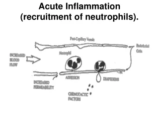
LEUKOCYTE EXTRAVASATION • Journey of leukocytes from the vessel lumen to the interstitial tissue, called extravasation.
LEUKOCYTE EXTRAVASATION-STEPS Across the endothelium In the lumen In interstitial tissue Margination Transmigration Rolling Chemotaxis Pavementing Adhesion to endothelium
Normally erythrocytes - central axial column, & leukocytes toward vessel wall. Cell free zone
In the lumen Margination Rolling Pavementing Adhesion to endothelium
Rolling/Activation Adhesion Transmigration Margination Selectins Integrins/Ig superfamily PECAM-1, others WBC Activation Endothelial Activation Chemoattractants Stimulus Chemotaxis
Leukocyte Extravasation-steps Margination • Process of leukocyte accumulation is called margination
Leukocyte Extravasation-steps Cell free zone
Rolling/Activation Adhesion Transmigration • Leukocytes tumble along the endothelium & adhere transiently (a process called rolling) Selectins Integrins/Ig superfamily PECAM-1, others WBC Activation Endothelial Activation Chemoattractants Rolling Stimulus Chemotaxis
Leukocyte Extravasation-steps Rolling Cell free zone • Leukocytes tumble along the endothelium & adhere transiently (a process called rolling)
Adhesion Rolling/Activation Adhesion Transmigration Selectins Integrins/Igsuperfamily PECAM-1, others WBC Activation Endothelial Activation Chemoattractants • Adhere firmly (resembling pebbles over which a stream runs without disturbing them). • Endothelium lined by white cells -called pavementing Stimulus Chemotaxis
Rolling/Activation Adhesion Transmigration Across the endothelium Selectins Integrins/Igsuperfamily PECAM-1, others WBC Activation Endothelial Activation Chemoattractants Escape into the extravascular space -Transmigration across the endothelium (also called diapedesis) Stimulus Chemotaxis
Rolling/Activation Adhesion Transmigration Across the endothelium Selectins Integrins/Igsuperfamily PECAM-1, others WBC Activation Insert pseudopods into the junctions between the endothelial cells, squeeze through interendothelial junctions, & assume a position between the endothelium & basement membrane Endothelial Activation Chemoattractants Stimulus Chemotaxis
Leukocyte Extravasation-steps Across the endothelium Escape into the extravascular space -Transmigration across the endothelium (also called diapedesis) Insert pseudopods into the junctions between the endothelial cells Blood vessel Squeeze through interendothelial junctions Opening of gap between endothelial cells
Rolling/Activation Adhesion Transmigration In interstitial tissue Chemotaxis WBC Activation Endothelial Activation Chemoattractants Stimulus Chemotaxis Migration in interstitial tissues toward a chemotactic stimulus
Leukocyte Extravasation-steps In interstitial tissue -Chemotaxis After extravasation, leukocytes migrate in the interstitial tissues toward the site of injury by a process called chemotaxis Chemotaxis=Defined most simply as locomotion oriented along a chemical gradient Stimulus
Phagocytosis and Clearance of the Offending Agent • Many leukocytes recognize, internalize, and digest foreignmaterial, microorganisms, or cellular debris by a processtermed phagocytosis. It consists of three steps • Recognition and attachment • Engulfment • Killing or degradation of the ingested materia
Recognition and Attachment • Receptors on the surface of phagocytic cells recognizecomponents of microbes and necrotic cells.
Recognition and Attachment • Leukocytes express several receptors that recognize external stimuli. These include • (1) receptors for microbial products (e.g. Toll-like receptors-TLRs) • (2) G protein–coupled receptors (recognize N-formylmethionie residues) • (3) receptors for cytokines (for INF-γ) and • (4) receptors for opsonins
PhagocytosisRecognition and Attachment • One of the main receptors is mannose receptors. • Phagocytosis is enhanced when microbes are opsonized • Microbes gets attached to the receptor Mannose receptor Microbe Lysosomes Leukocytes
Recognition and Attachment • Receptors for opsonins (phagocytic receptor): • The phagocytosis is enhanced when leukocyte receptorsrecognize microbes coated by specific host proteinsknown as opsonins. • The major opsonins are • IgG antibodies • C3b breakdown product of complement,certain plasma lectins called collectins
Clinical signifcance of opsonins • After exposure to antigen, B cells get activated and matureinto plasma cells, which produces immunoglobulins (IgG). • Bruton disease: Defect in maturation of the B-cells leadingto absence of immunoglobulin production. Hence, there isdefective opsonization.
Phagocytosis Engulfment. • Next step in phagocytosis is engulfment and formation of a phagocytic vacuole. • Phagocyte membrane zips around microbe. Mannose receptor Microbe Lysosomes
Phagocytosis Engulfment. • Phagosome: Extensions of the cytoplasm of leukocyteform pseudopods surrounding the particle to be ingested and forms a vesicle or vacuole called a phagosome. Mannose receptor Microbe Lysosomes
Engulfment Microbe ingested in phagosome Phagosome with ingested microbe Lysosome with enzymes
Engulfment Phagolysosome Membrane of phagosome fuses with membrane of a lysosomal granule
Engulfment Discharge of the granule's contents into the phagolysosome
Engulfment • Phagolysosome: The membrane of phagosome fuseswith membrane of lysosome to form a phagolysosome.Lysosomal granules are discharged into this phagolysosome
Clinical significance of defects in phagolysosomefunction • Chédiak-Higashi syndrome: Autosomal recessive conditioncharacterized by: • Increased susceptibility to infections: Due to defectivefusion of phagosomes and lysosomes in phagocytes.
Killing and Degradation • Killing and degradation of ingested microbial agents/particles occurs within neutrophils and macrophages. • Most important microbicidal agents are: • (1) reactive oxygen species • (2) reactive nitrogen species-derived from nitric oxide (NO) • (3) lysosomal enzymes.
Killing and Degradation • Reactive oxygen species (ROS): • Types of ROS are:
Reactive oxygen species (ROS) • Mechanism of production • In the phagocytic vacuole of leukocyte, rapid activation of NADPH oxidase (also called phagocyte oxidase), oxidizes NADPH (reduced nicotinamide-adenine dinucleotide phosphate) to NADP.
Reactive oxygen species (ROS): • Amount of H2O2 is insufficient to kill most of themicrobes by itself but the enzyme myeloperoxidase(MPO) present in the azurophilic granules of neutrophils can convert it into a powerful ROS. • MPO in the presence of a halide such as Cl–, converts H2O2 to hypochlorousradical (HOCl•), which is a potent oxidantand antimicrobial agent. • Hypochlorite (HOCl•) destroys microbes either by halogenation or by proteins and lipid peroxidation. • H2O2is also converted to hydroxyl radical (•OH) whichis also powerful destructive agent.
Reactive nitrogen species • NO, which is generated from arginine by the action of nitricoxide synthase (NOS), can kill microbes similar to ROS. • NO reacts with superoxide and produces highlyreactive free radical peroxynitrite (ONOO•).
Lysosomal enzymes • Acid hydrolases of lysosomes degrade the dead microorganisms. • Elastasecan kill bacteria.
Lysosomal enzymes • Constituents of leukocyte granules: The microbicidalsubstances within leukocyte cytoplasmic granulesinclude: • Bactericidal permeability—increasing protein • Lysozyme and lactoferrin • Major basic protein (MBP) present in eosinophils iscytotoxic to many parasites • Defensinsare toxic to microbes • Cathelicidins: These are antimicrobial proteins in theneutrophils and other cells. They are very effectiveagainst Mycobacterium tuberculosis