Download

1 / 25
260 likes | 612 Views
Acute cellular and humoral rejection. Eva Honsova Institute for Clinical and Experimental Medicine Prague, Czech Republic eva.honsova@ikem.cz.

E N D
Acute cellular and humoral rejection Eva Honsova Institute for Clinical and Experimental Medicine Prague, Czech Republic eva.honsova@ikem.cz
1.Cellular theory(Peter Medawar):the process of graft rejection is immunologically dependent.The cells(easy to see on histological slides)were recognized and identified as a cause of tissue damage. Graft rejection: The new era of transplantation began during World War II.
Hyperacute rejection (HR) • The antibodies are not seen in routine histological staining. • HR can occur immediately after transplantation • HR was observed when the kidney was transplanted into a recipient with pre-existing DSA • HR can also be caused by antibodiesdirected at allogenic blood groups A and B • New generation of laboratory tests prevents HR thrombosis
Graft rejection: • 2. Humoral theory (Paul Terasaki): evidence that alsoin later periods after transplantation antibodies must participate in graft damage. 2000: 80/70% at 5 years They were unable to prove this theory: no tissue marker of confirmation. Several landmark observations enabled the definition of dg. criteria of A-MR: P. Halloran: AR with „de novo“ DSAs production and poor prognosis. H. Feucht: studied complement components and recognized the relationship of C4d deposition in PTCs in kidney graftsto graft dysfunction.
Complement system C4d Positive C4d staining is a marker of antibody-mediated rejection and represents something like an imprint that the reaction occurred at the place of positive staining.
Detection of C4d (positive staining in PTCs) immunofluorescence immunoperoxidase
Classification of rejection changes • various centers were developing their own protocols and their own definitions of the morphological features • without standardization of the nomenclature the communication among various centers would remain unsatisfactory • Banff classification since 1991 • The schema underwent considerable evolution, and was repeatedly revised and modified in follow-up meetings during a 2-year period
Kidney graft biopsy, adequacy criteria • A large number of conditions can affect the allograft, frequently in combination with varying degrees of arteriosclerosis. • the pre-biopsy clinical diagnosis differed from the definitive diagnosis in 42%of episodes of graft dysfunction (Pascual et al. Transplantation 1999;67:737-41) • Therapy is changed in 60% following graft biopsy a) adequacy of the sample; b) processing of the tissue
The immunologic threat to the graft begins before transplantation A. Systemic effects of ischemia/reperfusion injury I/R up-regulates the expression of HLA antigens by the graft, the release of a cascade of chemokines, proinflammatory cytokines, and adhesion molecules. The increased display of HLA antigens intensifies the immune response. B. Each graft has its “medical history“; varying degrees of preexisting, clinically silent injury, mainly vascular nephrosclerosis No collateral vessel among arteries and arterioles; stenosis of the lumen represents chronic ischemia in the interstitial tissue.
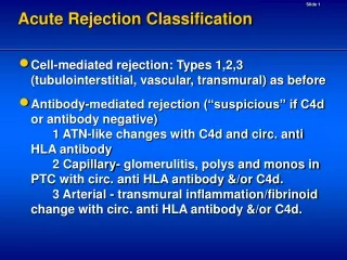
Acute T-cell-mediated rejection C: 3. Borderline Changes:"Suspicious" for acute T-cell-mediated r. C: 4. Acute T-cell-mediated rejection (type/grade) • IA.Cases with significant interstitial infiltration (>25% of parenchyma affected, i2 or i3) and foci of moderate tubulitis (t2) • IB. Cases with significant interstitial infiltration (> 25% of parenchyma affected, i2 or i3) and foci of severe tubulitis (t3) • IIA.Cases with mild to moderate intimal arteritis (v1, <25% of the luminal area) • IIB. Cases with severe intimal arteritis comprising > 25% of the luminal area (v2) • III. Cases with "transmural" arteritis and/or fibrinoid change and necrosis of medial smooth muscle cells with accompanying lymphocytic inflammation (v3)
Acute T-cell-mediated rejection, tubulitis-schema Significant lowering of the rate of episodes of ACR to approximately 5-10%in the 1st year Grade I: 65-70%, grade II: 30% Huge progress was made in our understanding of immunological mechanisms of rejection CD4 recognize peptide antigens presented by class II, can be divided according to their cytokine production: Th1: IFNγ , IL2, TNFα, typically in infiltrate of acute rejecting grafts TH2: IL4,IL5,IL10, provide help for B cells and production IgE – promote eosinophils Inflammatory cytokines produced by T cells activate epithelial cells, which in turn attract more T cells. CD8 recognize peptide antigens presented by class I MHC, produce IFNγ Mediate direct cytotoxicity via granzymes and perforin
Acute T-cell-mediated rejection, type I tubulitis in non-atrophic tubules Banff criteria: • IA. i:>25% of parenchyma affected, i2 or i3 and t2 • IB. i> 25% of parenchyma affected, i2 or i3 and t3 Chapel Hill Standards 1. i ≥5% 2. Mild to moderate interstitial edema 3. Tubulitis (t1 in ≥3 tubules in most inflamed area), - scattered eosinophils and ATI are common - MHC class II in tubular cells - If criteria 1-3 are not fulfilled, but tubules strongly expressed MHC class II, then an episode of ACR is suggested
Acute T-cell-mediated rejection, type Idg. problems and differential diagnosis • How much inflammation is reactive in subcapsular space early posttransplant • Resolving/partially treated rejection • inflammation in areas of interstitial fibrosis with t1 in the interface areas • 20-65% ATMR show C4d deposits along PTCs: mixed cellular- and antibody-mediated rejection Differential diagnosis • Drug-induced TIN Difficult or impossible to distinguish from ATMR • Pyelonephritis neutrophlic casts • Polyomavirus nephropathy plasma cells, IH detection
Acute T-cell-mediated rejection, type IDif. Dg.: polyoma virus nephropathy
Acute T-cell-mediated rejection, type II and IIIarteritis or endotheliitis
Acute T-cell-mediated rejection, type II and III differential diagnosis If we strictly follow the Banff criteria, one lymphocyte is enough for the dg. type II ATMR rejection. Pre-existing donor disease AS, hypertension Atheromatous embolism Severe hypertension
Acute B-cell-mediated rejection/humoral rejection • Recognition of AMR requires demonstration of C4d and circulating antibodies. The diagnostic criteria of AMR (cases that meet 2 criteria are considered suspicious for AHR) 1. Neutrophil/lymphocyte margination (capillaritis), and/or thombosis/necrosis 2. C4d+ in PTCs 3. Circulating anti-donor antibodies (DSA) Categories: 1.Hyperacute rejection 2. Acute B-cell-mediated rejection, late acute B-cell-mediated 3. Accommodation
Hyperacute rejection (HR) • Now, HR is not included in Banff schema • Complement dependent • recipient with pre-existing DSA • Can be C4d negative Differential dg.: • Vascular thrombosis (artery or vein), technical problems or hypercoagulable state • Donor TMA
Acute B-cell-mediated rejection/humoral rejection C4d is the terminal inactive split product of antibody-activated classical complement cascade and positive C4d staining represents something like animprint of the reactiondriven by antibodies. Scoring of C4d staining • C4d0 negative 0% of ptcs • C4d1 minimal C4d detection 1-10% • C4d2 focal C4d positive 11-50% • C4d3 diffuse C4d positive >50%
Acute B-cell-mediated rejectioncapillaritis: gli, PTCs; thrombosis AHR has occurred with all IS regiments. The pathology of AHR has a wide spectrum of findings. However, none of these features is specific.
Acute B-cell-mediated rejection Anti-donor Abs such as those directed at MHC antigens can trigger AMR. However, the absence of DSA cannot be taken to exclude AMR. Cases with: C4d+, DSA+ Cases with: C4d+, DSA- • some antigens may be expressed on the endothelial cells and not on lymphocytes, which are typically used for the test (MICA). • graft can absorb huge amounts of antibodies from blood and Abs can be below the level of detection. Cases with: C4d-,DSA+ • Negative staining may result from non-complementfixing antibodies, low reactivity of Abs. Sis B; Halloran P. Endothelial transcripts uncover a previously unknown phenotype: C4d-negative antibody-mediated rejection. Current Opinion in Organ Transplantation. 15(1):42-48, February 2010.
Acute B-cell-mediated rejection Phenomenon of so-called accommodation was repeatedly described in AB0 incompatible kidney grafts. In this situation, there are DSA, and C4d positive staining in PTCs. There is no tissue injury in the graft and the function is stable. However, in long-term studies, the morphological features of chronic injury were demonstrated more frequently than in AB0 compatible recipients. cg
Rejection remains a central challenge in the field of transplantation Although huge progress has been made in our understanding ofimmunological mechanisms of rejection there is still a lot unknown. ??? • Is humoral response always pathogenic? • What is the role of IgG subclasses in transplantation? • Can some antibodies block these DS antibodies which bind the complement? • Are all DSA harmful? • Is there acute antibody mediated rejection with arterial injury similar to type II ATMR? (humoral v, in mice it is so) (Hirohashi et al. Am J Transplant 2010:10;510-517)
Now we do not know what technique will represent a breakthrough and a great improvement in diagnosis of rejection; gene transcripts or perhaps microRNA, or something unknown … ???