1 / 0

Ankle problems/procedures and techniques

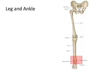
Ankle problems/procedures and techniques. Ankle Arthroscopy. Indications. Diagnostic Treatment Impingement Removal FB Stabilisation OCD’s Fusion procedures. Set up is everything. Mark out important structures like peroneal nerve Think about traction Inflate ankle with saline.

sofia
Download Presentation

Ankle problems/procedures and techniques

An Image/Link below is provided (as is) to download presentation Download Policy: Content on the Website is provided to you AS IS for your information and personal use and may not be sold / licensed / shared on other websites without getting consent from its author. Content is provided to you AS IS for your information and personal use only. Download presentation by click this link. While downloading, if for some reason you are not able to download a presentation, the publisher may have deleted the file from their server. During download, if you can't get a presentation, the file might be deleted by the publisher.

E N D

Presentation Transcript


  1. Ankle problems/procedures and techniques
  2. Ankle Arthroscopy

  3. Indications Diagnostic Treatment Impingement Removal FB Stabilisation OCD’s Fusion procedures
  4. Set up is everything Mark out important structures like peroneal nerve Think about traction Inflate ankle with saline
  5. Portals Anteromedial Medial to tib ant Anterolateral Either side of peroneal nerve Incise through skin only, use clip to bluntly get into ankle joint after inflating
  6. What to look for 8 point anterior examination Will need to switch camera from medial to lateral portal to see everything
  7. Ankle instability Physio is important to work on proprioception and peroneal recruitment and resolves most cases Seen in the NHS as a more chronic picture were ATFL +/- CFL stretched with resultant poor propriceptionperoneal recruitment
  8. Anatomy of lateral ligament complex
  9. Diagnosis of ligament injury
  10. Surgery for modified Brostrum Use longitudinal incision as also allows access to peroneal tendons Try and place anchors in position of ATFL and CFL
  11. Ankle fusion Can do anterior or lateral approach If lateral remove fibula (and can replace this if you want) Can use plate or screws Aim for neutral alignment with heel 5 degrees valgus Remove all cartillage. Make sure you have petallated bed of bleeding cancellous bone
  12. Open lateral approach with fibula osteotomyI Take off fibula just proximal to ankle Open ankle joint with Hinterman retractors Use lateral talar process as landmark for first wire and go distal to proximal
  13. Open lateral approach with fibula osteotomyII Once length measured drill wire more proximally out through medial skin and place first screw ( don’t spear yourself) Check on AP and LAT
  14. Post op for ankle fusion Non weight bearing 6 weeks in plaster Further 6 weeks weight bearing in aircast boot Physio at 8 weeks If smoker usually add on extra 4 weeks to above
  15. Arthroscopic ankle fusion Will need traction Make sure cartillage removed and subchondral bone exposed to allow to bleed Fix with 2 percutaneous screws
  16. Syndesmosis Injury 5-10% of ankle sprains Rupture of the interosseous ligaments between the tibia and fibula with or without fibular fracture Medial malleolar fracture or deltoid ligament rupture Persistent instability and gap in the joint after bimalleolar fixation
  17. Syndesmosis Exam Squeeze Test Squeeze the syndesmosis above the anklepain Abduction-External Rotation Stress Test Further instability with external rotation (may be shown with x-ray)
  18. Syndesmosis injuries AP/LAT/OBLIQUE ankle x-rays Syndesmotic widening Medial joint space widening Presence of fibula FX External rotation stress x-rays Severe pain associated with normal x-rays Must get tib/fib x-rays to rule out high fibula fracture
  19. Syndesmosis surgical management Gently dorsiflexhindfoot Internally rotate to align fibula with tibia Consider using large clamp if unable to reduce and internally rotate the fibula and compressing it to the tibia Perform medial arthrotomy if unable to reduce in order to debride medial ankle joint
  20. How big,how long and where to put it? 1 or 2: 3.5 or 4.5 mm cortical screws (no absolute evidence) 3 or 4 cortices (no evidence) Placed 1.5-2.0 cm superior to ankle joint line parallel to ankle joint from the posterolateral fibula to the anteromedial tibia Screws are not lagged! Posterior
  21. Tightrope for fixation Potentially better reduction of syndesmosis No need to remove Earlier weight bearing
More Related