Download
1 / 37
380 likes | 755 Views
Foot and Ankle II. RF/Subtalar Joint Varus – Compensated ST Varus & FF Valgus – Flexible PF 1 st Ray. RF/Subtalar Joint Varus - Pathomechanics. Varus position of RF @ IC magnitude of pronation duration of pronation rapid supination following TO. RF/Subtalar Joint Varus - Compensation.

E N D
RF/Subtalar Joint Varus – Compensated ST Varus & FF Valgus – Flexible PF 1st Ray
RF/Subtalar Joint Varus - Pathomechanics • Varus position of RF @ IC • magnitude of pronation • duration of pronation • rapid supination following TO
RF/Subtalar Joint Varus - Compensation • Excessive pronation at STJ
RF/Subtalar Joint Varus – Compensated ST Varus & FF Valgus – Flexible PF 1st Ray
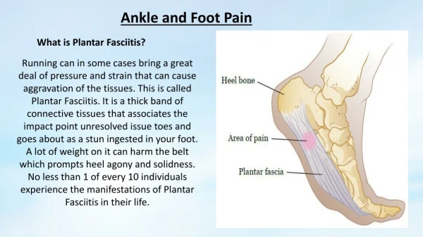
RF/Subtalar Joint Varus - Pathology • Chronic ankle sprains 20 calcaneal EV • Plantar keratosis @ 2nd MET • PL tendinitis 20 rapid supination • TP tendinitis 20 deceleration of pronation • Achilles tendinitis 20 active supination
Supination Closed-chain • Calcaneus inversion (varus) • Talus abduction (ER - vertical axis) • Talus dorsiflexion • Tibial ER
Abnormal Supination • Excessive in magnitude • Excessive in duration • Occurs at wrong time Causes: • Intrinsic deformities • Extrinsic deformities
Abnormal Supination - Etiology • FF valgus • PF first ray • FF equinus deformities
Abnormal Supination - Pathology • Lack of shock absorption • plantar lesions • 1st and 5th ray trauma • abnormal pronation during propulsion
Forefoot Valgus - Rigid PF 1st Ray/Total • 1st ray always p-flexed relative to other MET heads • medial foot load prematurely • lateral aspect of foot loaded prior to HO • “abnormal supination”
Peroneus Longus Pulley • lateral malleolus • calcaneus - peroneal notch • cuboid - peronal groove • base of 1st MET and medial cuneiform
Peroneus Longus Pulley • Pronated Foot
Peroneus Longus Pulley • plantarflexor of 1st ray • cinches tarsal-metatarsal articulations • FF --> HO
Tibialis Posterior • distal lateral tibia • navicular • attaches to all tarsals (except talus) • attaches to base of MET 2-4
Tibialis Posterior • Strong supinator • HS --> FF • late midstance --> HO • inconsistent thru MSt IC LR MSt TSt PSw ISw MSw TSw IC
FHL • medial malleolus • talus • 1st MET head (sesamoid) • attaches to distal phalanx • cinches foot • assists w/ supination • FF --> TO
FHL • cinches foot • assists w/ supination • MSt --> TO IC LR MSt TSt PSw ISw MSw TSw IC
Conclusions • Pronation - hypermobilities • Supination - hypomobilities Either can cause a reduced ability to: • attenuate forces • convert torque • adapt to terrain • become a rigid lever
Guidelines for Posting Maximal FF posting: • males: 7 - 8 mm (10 = 1 mm) • females: 5 - 6 mm • shoewear dependent
Guidelines for Posting FF Varus Deformity: • medial FF area • If FF deformity > maximal FF posting allows post RF 4mm) FF Valgus Deformity: • lateral FF area
Guidelines for Posting Equinus Deformity: • stretching w/ foot in STJN • lift RF 50% of lacking range - maybe done initially in acute cases • maximum in-shoe lift: 0.25” (7mm) • balance out contralateral limb