Download
1 / 48
610 likes | 1.92k Views
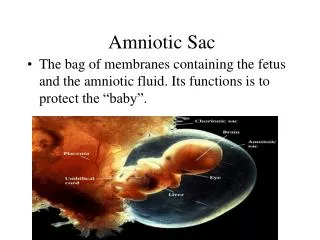
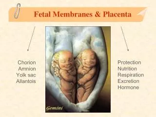
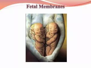
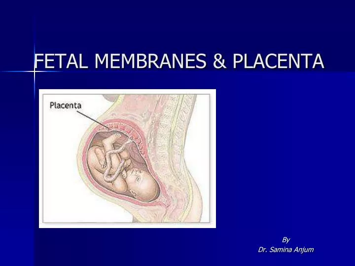
FETAL MEMBRANES & PLACENTA. By Dr. Samina Anjum. FETAL MEMBRANES. In vertebrate embryo only part of egg forms the actual embryo while other part is known as the extra embryonic and forms fetal membranes. Fetal membranes consist of: Yolk Sac Amnion Chorion Allantois Placenta.

E N D
FETAL MEMBRANES & PLACENTA By Dr. SaminaAnjum
FETAL MEMBRANES In vertebrate embryo only part of egg forms the actual embryo while other part is known as the extra embryonic and forms fetal membranes. Fetal membranes consist of: • Yolk Sac • Amnion • Chorion • Allantois • Placenta
PLACENTA • Is an organ that facilitates nutrient and gas exchange b/w maternal and fetal compartments • As the fetus begins the 9th week of development , its demand for nutritional and other factors increases, causing major changes in placenta.
DEVELOPMENT OF TROPHOBLAST By beginning of third week DEVELOPMENT OF A VILLUS
Outer cytotrophoblast shell. Stem (anchoring) villi Free (terminal) villi.
TROPHOBLAST AT THE END OF THIRD WEEK OF DEVELOPMENT • When heart begins to beat in the 4th week, villous system is ready. • Intervillous spaces lined with syncytiotrophoblast
CHANGES IN TROPHOBLAST • Placenta consists of two components: • Fetal portion is derived from trophoblast and extraembryonic mesoderm • Maternalportion is derived from uterine endometrium
TROPHOBLAST AT THE BEGININING OF 2ND MONTH OF DEVELOPMENT • Radial appearance of trophoblast due to increased no. of Secondary & Tertiary villi • Villi cover the entire surface of chorion
FORMATION OF HYBRID VESSELS • Endovascular invasion • Epithelial to endothelial transition • Transformation of spiral arteries from small diameter, high resistance vessels to larger diameter, low resistance vessels • Hybrid vessels form
STRUCTURE OF VILLI AT DIFFERENT STAGES OF DEVELOPMENT Syncytial knots Anchoring villi Free villi 4th week 4th month
STRUCTURE OF CHORION Difference b/w embryonic and abembryonic poles of chorion • Chorion frondosum • Chorion laeve
STRUCTURE OF DECIDUA(GRAVID ENDOMETRIUM) This difference b/w embryonic and abembryonic poles is also reflected in the structure of decidua Decidua(that which falls off) is the functional layer of endometrium which is shed during parturition.
PARTS OF DECIDUA • Deciduabasalis(deep to the conceptus/underlying the conceptus or b/w it and the muscular wall of uterus). • Deciduacapsularis (overlying the conceptus/covering the chorionic sac and interposed b/w sac and uterine cavity). • Deciduaparietalis(remaining lining of uterine endometrium exclusive of the area occupied by embryo)
With growth of chorionic vesicle, D.Cap. is stretched & degenerates • Chorion leave then comes in contact with uterine wall (the Decidua Parietalis) and two fuse and obliterates the uterine lumen. • Fusion of amnion and chorion together form amniochorionic membrane which ruptures during labour
STRUCTURE OF PLACENTA • Placenta consists of two components. • Fetal portion (Chorion Frondosum) • Maternal portion (Deciduabasalis)
Fetal side placenta is bordered by chorionic plate • Maternal side is bordered by deciduabasalis. • Junctional zone: Trophoblast & maternal cells intermingle • Intervillous space: b/w decidual plate & chorionic plate
During 4th to 5th monthdecidual septaproject into intervillous spaces but do not reach chorionic plate. • Septa have a core of maternal tissue but surface is covered by syncytial cells which separates the maternal blood from fetal tissue of villi.
Placenta is divided into cotyledons by septa however contact between them is maintained. • Placenta enlarge with advancement of pregnancy and may occupy 15 to 30% of uterine space.
FULL TERM PLACENTA • Discoid in shape • Diameter 15-25cm • 3cm thick • Weight 500-600g • Expulsion about 30 minutes after child birth
No. of cotyledons 15-20 visible on maternal side after child birth. • Cotyledons are covered by thin layer of decidua basalis.
Fetal surface of placenta is covered by chorionic plate; Chorionic vessels converge towards umbilical cord • Amnion • Attachment of umbilical cord is eccentric
CIRCULATION OF THE PLACENTA • Cotyledons receive their blood through 80-100 spiral arteries • Collectively the Intervillous spaces of a mature placenta contain approx 150 ml of blood which is replenished 3-4 times / min • This blood moves along chorionic villi which have a surface area of 4-14 meter square
CLINICAL CORRELATES • Erythroblastosisfetalis • Synthetic progestinsmasculanize female fetuses • Diethylstilbestrol can cause CA of vagina • Fetal immunity is provided against diphtheria, small pox and measles but not against pertussis, and varicella (chicken pox)
Types of placenta • One classification scheme for placentas is based on which maternal layers are retained in the placenta or which maternal tissue is in contact with chorionic epithelium of the fetus.
Type of Placenta Maternal Layers Retained
Types of placenta • Placenta Accreta: An invasion of the myometrium which does not penetrate the entire thickness of the muscle. • Placenta Increta: Occurs when the placenta further extends into the myometrium. • Placenta Percreta: The worst form of the condition is when the placenta penetrates the entire myometrium to the uterine serosa (invades through entire uterine wall). This variant can lead to the placenta attaching to other organs such as the rectum or bladder • Placenta previa: Blastocyct implants close to or overlying the internal os of uterus.
AMNION AND UMBILICAL CORD • The oval line of reflection between amnion & ectoderm is amnio-ectodermal junction (primitive umbilical ring). • Following structures pass through the ring at the 5th week of development: • Connecting stalk,containing Allantois and the umbilical vessels • Yolk stalk (vitelline duct), accompanied by vitelline vessels • Canal connecting the intraembryonic and extraembryonic cavities
10th week 5th week • The embryonic cavity enlarges rapidly and the amnion envelops the connecting and yolk sac stalks crowding them and giving rise to primitive umbilical cord. • Formation of physiological umbilical hernia. • At the end of 3rd month loops with draw, chorionic cavity and yolk sac are obliterated. • When the canal, allantois and the vitelline duct and its vessels are also obliterated, all that remains in the cord are the umbilical vessels surrounded by the jelly of Wharton. Wharton’s jelly is rich in proteoglycans, functions as a protective layer for blood vessels. The walls of the vessels are muscular and elastic, once the cord is tied off, rapidly constrict.
Umbilical cord (2cm thick ; 50-60cm long) • Battledore placenta: Marginal insertion • Velamentous insertion of cord: attached to fetal membranes
PLACENTAL CHANGES AT THE END OF PREGNANCY • Increase in fibrous tissue in the villus core • Thickening of basement membranes in fetal capillaries • Obliterative changes in small capillaries of villi • Fibrinoid deposition on the surface of villi (infarction of intervillous lake or of entire cotyledon giving whitish appearance)
AMNIOTIC FLUID • Amniotic cavity is filled with clear watery fluid • Derived from maternal blood and amniotic cells • 800-1000ml at 37 weeks • By fifth month fetus swallows own amniotic fluid- about 400ml a day • Fetal urine is added daily in amniotic fluid in 5th month
Functions of Amniotic Fluid • Absorb jolts • Prevent adherence to the amnion • Allows free fetal movements • Helps in dilatation of cervical canal at the time of birth
Clinical correlates Polyhydroamnios(more than1500-2000 ml) • Causes: Idiopathic, maternal diabetes, congenital malformations, anencephaly, esophageal atresias. Oligohydroamnios(less than 400 ml) • Causes: Renal agenesis, premature rupture of membranes.
FETAL MEMBRANES IN TWINS • Dizygotic/Fraternal twins (womb- mates)2/3rd • 7-11 / 1000 births • Simultaneous shedding of two oocytes and fertilization by different spermatozoa • Hereditary tendency • Incidence increases with maternal age • Sex may be same or opposite • Resemblance like brothers and sisters • Zygotes implant individually in the uterus and each develops its own placenta, amnion, and chorionic sac • Erythrocyte mosaicism
Monozygotic twins (identical twins) • Develop from single fertilized ovum • 3-4/1000 births • Genetically identical • Result from splitting of zygote at an early stage • Close resemblance in blood groups, fingerprints, sex and external appearance such as eye and hair color
TWIN DEFECTS • Erythrocyte mosaicism: Anastomosis b/w blood vessels of fused placenta of DZ; RBCs of two different types. • Twin transfusion syndrome: Shunting of arterial blood from one twin through AV anastomosis into venous circulation of other twin. Donar twin is anemic while the recipient twin is polycythemic. • Fetus papyraceous: Death and resorption of one fetus
CONJOINED OR SIAMESE TWINS(Partial splitting of primitive node & streak)
PRETERM BIRTH (Delivery before 34 weeks) Causes: • Premature rupture of membranes • Premature onset of labor • Pregnancy complications requiring premature delivery • Maternal hypertension • Maternal Diabetes • Maternal vaginal infections