Download
1 / 42
430 likes | 776 Views
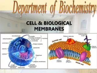
MEMBRANES Membranes are sheets of tissues that cover or line surfaces or that separate organs or parts(lobes) of organs from one another. . Types of membranes: 1- Epithelial membranes 2- Connective tissue membranes. EPITHELIAL MEMBRANES. two types of epithelial membranes: serous

E N D
MEMBRANESMembranes are sheets of tissues that cover or line surfacesor that separate organs or parts(lobes) of organs from one another. Types of membranes: 1-Epithelial membranes 2-Connectivetissue membranes
EPITHELIAL MEMBRANES • two types of epithelial membranes: • serous • and mucous. • Each type is found in specific locations within the body and secretes a fluid. *These fluids are called -serous fluid -and mucus.
Mucous Membranes • Mucous membranes line the body systems that have openings to the environment. (respiratory, digestive, urinary, and reproductive). • The epithelium of a mucous membrane (mucosa) varies: - mucosa of the esophagus and of the vagina is stratified squamous • mucosa of the trachea is ciliated epithelium • mucosa of the stomach is columnar epithelium
Mucous Membranes • Mucosa is composed of epithelium resting on a loose connective tissue membrane called a lamina propria • The mucus secreted keeps the lining epithelial cells wet and acts as lubricant
Serous Membranes • Serous membranes are sheets of simple squamous epithelium that line some closed body cavities and cover the organs in these cavities • 1-pleural membranes are the serous membranes of the thoracic cavity. • The parietal pleura lines the chest wall. • and the visceral pleura covers the lungs. • It secrete serous fluid, which prevents friction between them
2-pericardium • The heart, has its own set of serous membranes. • The parietal pericardiumlines the thorasic cavity • the visceral pericardium, or epicardium, is on the surface of the heart muscle • Serous fluid is produced to prevent friction as the heart beat
CONNECTIVE TISSUE MEMBRANES • These are made of connective tissue with no epithelial cells at all • It cushions organs moving against each other during muscle activity
The Integumentary System • consists of : -the skin, - its accessory structures such as hair and sweat glands, -and the subcutaneous tissue below the skin. * The skin is considered an organ.
Functions of the skin 1-Protects deeper tissues from: • -mechanical damage • -chemical damage (acids & bases) • -bacterial damage • -ultraviolet radiation (sunlight) • Thermal (heat or cold) damage • -desiccation (drying out)
Functions of the skin (cont.) 2-aids in heat regulation -in cold: vasogonstriction -in hot: vasodilatation+sweat 3-aids in excretion of urea & uric acid 4-synthesize vitamin D
STRUCTURES OF THE SKIN • two major layers the epidermis &dermis. • The epidermis: • made of stratified squamous keratinizing epithelial tissue and is thickest on the palms and soles. The cells that are most abundant are called keratinocytes, and there are no capillaries present between them.
The epidermis: • Although the epidermis may be further • subdivided into four or five sublayers, two of these are of greatest importance: • the innermost layer, the stratum germinativum, • and the outermost layer, the stratum corneum .
Stratum Germinativum(BASALE) • To germinate means “to sprout” or “to grow.” Basal means the “base” or “lowest part.” The stratum germinativum is the base of the epidermis, the innermost layer in which mitosis takes place. New cells are continually being produced, pushing the older cells toward the skin surface. These cells produce the protein keratin, and as they get farther away from the capillaries in the dermis, they die.
Stratum Corneum • the outermost epidermal layer, • consists of many layers of dead cells; all that is left is their keratin which is a fibrous protein that makes the epidermis: - a tough protective layer - relatively waterproof (prevent evaporation, prevents the entry of water). -a barrier to pathogens and chemicals. * Stratum Corneum rubs and flakes slowly leading to totally new epidermis every25-45 days
Langerhans Cells • are also called dendritic cells(branched appearance ) .These cells originate in the red bone marrow, and are quite mobile. They are able to phagocytize foreign material, such as bacteria that enter the body through breaks in the skin. With such ingested pathogens, the Langerhans cells migrate to lymph nodes and present the pathogen to lymphocytes. This triggers an immune response such as the production of antibodies.
Melanocytes • Another type of cell found in the lower epidermis • Melanocytes produce another protein, a pigment called melanin. • In people with dark skin, the melanocytes continuously produce large amounts of melanin. The melanocytes of lightskinned people produce less melanin. The activity of melanocytes is genetically regulated.
Melanin • In all people, melanin production is increased by exposure of the skin to ultraviolet rays, which are part of sunlight and are damaging to living cells. • People with dark skin already have good protection against the damaging effects of ultraviolet rays. • Melanin also gives color to hair,iris &choroid.
DERMIS • It is a strong stretchy envelope that hold the body together • It is made of an irregular type of fibrous connective tissue. • . Fibroblasts produce both collagen and elastin fibers. • Collagen fibers are strong, and elastin fibers are able to recoil after being stretched. • Strength and elasticity are two characteristics of the dermis. • With increasing age, however, the deterioration of the elastin fibers causes the skin to lose its elasticity.
papillary layer of dermis • IT is the uneven junction of the dermis with the epidermis • Capillaries are abundant here to nourish not only the dermis but also the stratum germinativum. N.B. The epidermis has no capillaries of its own. • Within the dermis are the accessory skin structures: hairand nail follicles, sensory receptors, and several types ofglands.
Colour of skin • Three pigments contribute to skin golour • 1-melanin • 2-karotine • 3-oxyhemoglobin
Appendages of the skin • 1-Hair follicles—mitosis takes place in the hair root; new cells produce keratin, die, and become the hair shaft. • Hair of the scalp provides insulation from cold for the head; eyelashes keep dust out of eyes; • nostril hairs keep dust out of nasal cavities
2. Nail follicles—at the ends of fingers and toes; mitosis takes place in the nail root; the nail itself is dead,keratinized cells. -Nails protect the ends of the fingers and toes, enable the fingers to pick up small objects, and provide for efficient scratching
3. Receptors—detect changes in the skin: touch, pressure, heat, cold, and pain; provide information about the external environment that initiates appropriate responses. • sensitivity of the skin depends on the number of receptors present.
4. Sebaceous glands—(oil glands) secrete sebum into hair follicles or to the skin surface. • sebum inhibits the growth of bacteria and prevents drying of skin and hair. • If its duct is blocked by sebum,whitehead appears,with oxidation become blackhead • Active infection leads to acne
5. Ceruminous glands—secrete cerumen in the ear canals. • cerumen prevents drying of the eardrum.
6. Apocrine sweat glands—modified scent glands in axillae and genital area; • activated by stress and emotions • Their secretions contains fatty acids and proteins • Secretion is odorless • Begins function at puberty • Minimal role in thermoregulation
7. Eccrine sweat glands—all over the body, most numerous on face, palms, soles. • Activated by high external temperature or exercise; sweat on skin surface is evaporated by excess body heat. Excretion of small amounts of NaCl and urea is a very minor function.
8. Arterioles—smooth muscle permits constriction or dilation. • Vasoconstriction in cold temperatures decreases dermal blood flow to conserve heat in the body core. • Vasodilation in warm temperatures increases dermal blood flow to bring heat to the surface to be lost. • Vasoconstriction during stress shunts blood away from the skin to more vital organs, such as muscles, to permit a physical response, if necessary.
Subcutaneous Tissue • Subcutaneous Tissue—also called the superficial fascia; connects skin to muscles • 1. Areolar tissue, called loose connective tissue; the matrix contains tissue fluid and WBCs that destroy pathogens • 2. Adipose tissue stores fat as potential energy; cushions bony prominences; provides some insulation from cold.
AGING AND THEINTEGUMENTARY SYSTEM • become thinner as mitosis slows and fibroblasts die and are not replaced; repair is slower. • becomes wrinkled as collagen and elastin fibers deteriorate. - Sebaceous glands and sweat glands become less active; the skin becomes dry, and temperature regulation becomes more difficult. • Hair follicles become inactive and hair thins. • Melanocytes die so white hair • Less fat in subcutaneous tissue so more sensitivity to cold