Download
1 / 11
110 likes | 123 Views
Microanalysis in Science and Engineering – Microscopy of Biological Samples. A Workshop for Middle and High School Teachers sponsored by Tennessee Technological University Center for Manufacturing Research Departments of Chemical, Mechanical, Earth Sciences and Curriculum and Instruction

E N D
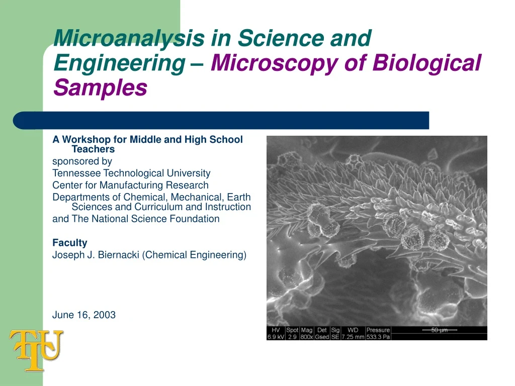
Microanalysis in Science and Engineering – Microscopy of Biological Samples A Workshop for Middle and High School Teachers sponsored by Tennessee Technological University Center for Manufacturing Research Departments of Chemical, Mechanical, Earth Sciences and Curriculum and Instruction and The National Science Foundation Faculty Joseph J. Biernacki (Chemical Engineering) June 16, 2003
What will we learn? • Why are biological samples different? • What can be done to make it possible to look at biological samples in the SEM? • What linkages can be made between the “technology fundamentals” and the middle/high school science curriculum?
Why are biological samples different? • Most biological and even many inorganic samples contain water or other volatile substances. These volatiles are typically incompatible with the high vacuum environment typically required by conventional SEM. A bit of history: Thornley (1960) – described a way to image liquid droplets in the SEM. Robinson (1974) – first laboratory full chamber atmosphere control. JEOL (1978) – first commercial variable pressure SEM (VPSEM). Electroscan (1987) – first differentially pumped environmental SEM (ESEM).
What can be done? • There are two options: • In some cases the water can be removed in such a way that the sample microstructure is somewhat retained. • A variable pressure or environmental SEM can be used which permits some level of gaseous environment to exist in the sample chamber. Such instruments are referred to as variable pressure or environmental SEMs depending on the pressure achievable.
Suggested Curriculum Links Chemistry: vapor pressure, boiling point, partial pressure So what is the issue with wet samples? • Water, for example, will boil at 23.7 torr (.031 atm) at 25 oC. Since an ordinary SEM operates at 10-5 torr, unprotected condensed water is simply incompatible. Even the vapor pressure of ice at 0 oC is 5 torr. • Electrons interact with gas in the same way they interact with solids only the frequency of collision is lower. • Gas molecules scatter electrons and therefore the effective spot size is altered due to “beam skirting.” This lowers resolution at otherwise same beam operating conditions.
Suggested Curriculum Links Chemistry: kinetic theory of gasses Interaction of e- with gas • The mean free path of an e- is a function of the gas pressure.
Suggested Curriculum Links Chemistry: surface tension What can be done to protect the sample? • If a conventional high vacuum SEM is used, the wet sample must be dehydrated. For inorganic samples this typically means drying at some elevated temperature to remove water. For biological sample, and even some inorganic materials, a process called critical point drying is usually used.
Suggested Curriculum Links Chemistry: critical point, surface tension, PVT relationship Critical point drying • Carbon dioxide (CO2) is typically used as the fluid in critical point drying. The water in the sample is replaced with an intermediate solvent, i.e. acetone, which in turn is dissolved by supercritical CO2. The conditions, T and P, are controlled in such a way that the CO2 can be evacuated from the system without going below the critical point. This prevents the formation of a gas-liquid interface thereby avoiding surface tension induced forces which are characteristic of evaporation of liquids from solids. http://www.emsdiasum.com/ems/equipment/critical_drying.html
Variable pressure and environmental SEM • When a significant amount of gas is present (1-20 torr) in the sample chamber there are options which permit electron detection. • Since backscattered electrons are very high in energy, they can be detected by conventional large-field detectors. • Gaseous secondary detectors have also been developed.
+ V - sample Gaseous secondary electron detection • Gaseous secondary detection utilizes amplification of secondary electrons by crating an ionization cascade. http://www.calce.umd.edu/general/Facilities/ESEM.pdf http://www.shef.ac.uk/uni/academic/N-Q/phys/teaching/phy311/gaseous.html
Summary • Electrons quickly loose their energy due to inelastic collisions with gas molecules. • Biological samples must be dried prior to viewing in a conventional SEM. • Environmental SEMs have special secondary detectors which are compatible with limited vapor pressures of gas up to about 20 torr.