Download
1 / 18
210 likes | 584 Views
“ Scanning Tunneling Microscopy Transmission Electron Microscopy”. Speakers Burcu Başar Semih Gezgin Yavuz Selim Telis Place Hacettepe University Department of Chemical Engineering

E N D
“Scanning Tunneling MicroscopyTransmission Electron Microscopy” Speakers Burcu Başar Semih Gezgin Yavuz Selim Telis Place Hacettepe University Department of Chemical Engineering “It’s a small world after all.. “ " April 12/2012
IN THIS PRESENTATION... Invention General Overview Basic Set-up How They Work Advantages&Limitations Applications Conclusion
INVENTION.. • STM -Invented by Binnig and Rohrer at IBM in 1981 (Nobel Prize in Physics in 1986) • TEM-Invented by Ernst Ruska and Max Knoll in 1931
GENERAL OVERVIEW.. STM • An electron microscope that uses a single atom tip to attain atomic resolution. • Topographic (real space) images • Spectroscopic (electronic structure, density of states) images TEM • Used to characterize the microstructure of materials with very high spatial resolution • Examine internal morphology of polymers from segmental to atomic level(e.g.block copolymers,crystalline polymers..) Iron atoms on the surface ofCu(111)[2]
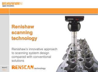
BASIC SET-UP.. STM includes; Scanning tip Piezoelectriccontrolledscanner Distance control and scanning unit Vibrationisolationsystem Computer TEM includes; Electrongun Condensersystem Specimenchamber Objective lens systems Projector lens systems
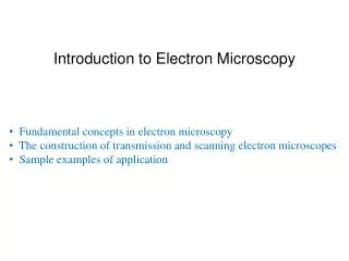
HOW TO OPERATE TEM?.. • Beam of electrons transmitted through an ultra thin specimen • Image formed, magnified and detected through a sensor • Image contrast produced through electron scattering by the atomic nuclei of the sample • Contrast within the sample enhanced by the use of stains
LIMITATIONS .. More time for sample preparation Require experience than most other techniques Samples damaged by the electron beam irradiation;having to withstand high vaccum The entire specimen fit into a 3mm diameter cup&Be less than 100 microns in thickness SBS block copolymer structure[3] Typical TEM image of a SBS sample
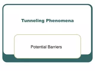
HOW TO OPERATE STM?.. “I think I can safely say that nobody understands QuantumMechanics.” Richard Feynman
How tunneling works ? Simple answer
What an STM measures?------Local density of states Each plane represents a different value of the tip-sample V, and thelateral position on the plane gives the x,y position of the tip. Filled states are given in red. The plane at the Fermi energy (V=0) is shown in blue.
ADVANTAGES.. • Conceptually simple but complexities in use • Can even move atoms • Can be used in variety of temperatures • Perform in different environments(air, water etc.)
LIMITATIONS.. • Slower compared to other techniques • Mainly used to analyze conducting materials • The best results from STM can be obtained only in vacuum conditions,hence it may not be the best tool to inspect and analyse biological samples Damaged tip[6]
WIDE USAGE AREA IN.. • Physics, semiconductor physics and microelectronics • Chemistry, surface reaction catalysis • Biology,in the study of DNA molecules • Nanoscale chemistry labs, synthetic chemical compounds
CONCLUDINGREMARKS.. STM vsTEM • Maintains a constant tunnelling electrical current • Very high resolution • Better resolution but limited to conducting materials • Offer the most powerful magnification, potentially over one million times or more • Ability to utilize in a variety of different scientific, educational and industrial fields • Provide information on element and compound structure • Easy to operate with proper training
REFERENCES.. • [1] (a)G. Binnig and H. Rohrer, U.S. Patent No. 4,343,993 (10 August 1982). (b)Binnig, G., Rohrer, H., et al., (1982) Phys. Rev. Lett., 49:57. • [2] G. Binnig, et al., Phys. Rev. Lett., 56, 930-933 (1986). • [3] The Tunneling Current - A Simple Theory http://wwwex.physik.uni-ulm.de/lehre/methmikr/buch/node5.html • [4] Scanning Tunneling Microscopy http://www.physnet.uni-hamburg.de/home/vms/pascal/stm.htm • [5] Scanning Tunneling Microscopy Basics http://nanowiz.tripod.com/stmbasic/stmbasic.htm • [6] Scanning Tunneling Microscopy http://www.chembio.uoguelph.ca/thomas/stm_research.html • [7] Interpretation of Scanning Tunneling Microscopy and Spectroscopy of Magnetic Metal Surfaces by Electron Theory, Daniel Wortmann,,Universityat Dortmund, Februar 2000,available online. • [8] Davis Baird,Ashley Shew,Department of Philosophy, University of South Carolina, Columbia, Probing the History of Scanning Tunneling Microscopy, October 2002,available online. • [9] Lecture 4,Scanning Tunneling Microscopy, CHM8490/8190, Spring 2000, Dr. Gang-yu Liu
“The reason we are on an imaginative level is not because we have finer imagination,but because we have better instruments.” Alfred North Whitehead THANK S FOR YOUR ATTENTION..