Download
1 / 17
170 likes | 331 Views
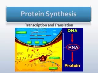
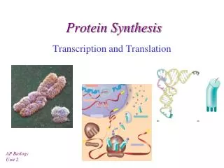
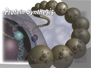
Protein Synthesis. Chapter 3, Section 6. Protein Synthesis. DNA is the master blueprint for protein synthesis Gene: Segment of DNA with blueprint for one polypeptide Triplets of nucleotide bases form genetic library Each triplet specifies coding for an amino acid. Nuclear envelope. DNA.

E N D
Protein Synthesis Chapter 3, Section 6
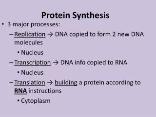
Protein Synthesis • DNA is the master blueprint for protein synthesis • Gene: Segment of DNA with blueprint for one polypeptide • Triplets of nucleotide bases form genetic library • Each triplet specifies coding for an amino acid
Nuclear envelope DNA Transcription RNA Processing Pre-mRNA mRNA Nuclear pores Ribosome Translation Polypeptide Figure 3.34
Roles of the Three Main Types of RNA • Messenger RNA (mRNA) • Carries instructions for building a polypeptide, from gene in DNA to ribosomes in cytoplasm
Roles of the Three Main Types of RNA • Ribosomal RNA (rRNA) • A structural component of ribosomes that, along with tRNA, helps translate message from mRNA
Roles of the Three Main Types of RNA • Transfer RNAs (tRNAs) • Bind to amino acids and pair with bases of codons of mRNA at ribosome to begin process of protein synthesis
Transcription • Transfers DNA gene base sequence to a complementary base sequence of an mRNA • Transcription factor • Loosens histones from DNA in area to be transcribed • Binds to promoter, a DNA sequence specifying start site of gene to be transcribed • Mediates the binding of RNA polymerase to promoter
Transcription • RNA polymerase • Enzyme that oversees synthesis of mRNA • Unwinds DNA template • Adds complementary RNA nucleotides on DNA template and joins them together • Stops when it reaches termination signal • mRNA pulls off the DNA template, is further processed by enzymes, and enters cytosol
RNA polymerase Coding strand DNA Terminationsignal Promoterregion Template strand 1 Initiation: With the help of transcription factors, RNApolymerase binds to the promoter, pries apart the two DNA strands,and initiates mRNA synthesis at the start point on the template strand. Template strand mRNA Coding strand of DNA Rewindingof DNA Unwindingof DNA 2 Elongation: As the RNA polymerase moves along the templatestrand, elongating the mRNA transcript one base at a time, it unwindsthe DNA double helix before it and rewinds the double helix behind it. RNA nucleotides Direction oftranscription Templatestrand mRNA transcript DNA-RNA hybrid region mRNA RNApolymerase 3 Termination: mRNA synthesis ends when the termination signalis reached. RNA polymerase and the completed mRNA transcript arereleased. The DNA-RNA hybrid: At any given moment, 16–18 base pairs ofDNA are unwound and the most recently made RNA is still bound toDNA. This small region is called the DNA-RNA hybrid. Completed mRNA transcript RNA polymerase Figure 3.35
Translation • Converts base sequence of nucleic acids into the amino acid sequence of proteins • Involves mRNAs, tRNAs, and rRNAs
Genetic Code • Each three-base sequence on DNA is represented by a codon • Codon—complementary three-base sequence on mRNA
SECOND BASE U C A G U UUU UCU UAU UGU Tyr Cys Phe C UUC UCC UAC UGC U Ser A UUA UCA UAA Stop UGA Stop Leu G UUG UCG UAG Stop UGG Trp U CUU CCU CAU CGU His C CUC CCC CAC CGC C Leu Pro Arg A CUA CCA CAA CGA Gln G CUG CCG CAG CGG U AUU ACU AAU AGU Asn Ser C Ile AUC ACC AAC AGC A Thr A AUA ACA AAA AGA Lys Arg Met or G AUG ACG AAG AGG Start U GUU GCU GAU GGU Asp C GUC GCC GAC GGC G Val Ala Gly A GUA GCA GAA GGA Glu G GUG GCG GAG GGG Figure 3.36
Translation • mRNA attaches to a small ribosomal subunit that moves along the mRNA to the start codon • Large ribosomal unit attaches, forming a functional ribosome • Anticodon of a tRNA binds to its complementary codon and adds its amino acid to the forming protein chain • New amino acids are added by other tRNAs as ribosome moves along rRNA, until stop codon is reached
Nucleus Energized by ATP, the correct amino acid is attached to each species of tRNA by aminoacyl-tRNA synthetase enzyme. RNA polymerase mRNA Leu Template strand of DNA Amino acid 1 After mRNA synthesis in the nucleus, mRNA leaves the nucleus and attaches to a ribosome. Nuclear pore tRNA Nuclear membrane A G A 2 Translation begins as incoming aminoacyl-tRNA recognizes the complementary codon calling for it at the A site on the ribosome. It hydrogen-bonds to the codon via its anticodon. Released mRNA Aminoacyl-tRNA synthetase Leu 3 As the ribosome moves along the mRNA, and each codon is read in sequence, a new amino acid is added to the growing protein chain and the tRNA in the A site is translocated to the P site. Ile tRNA “head” bearing anticodon G A A Pro 4 Once its amino acid is released from the P site, tRNA is ratcheted to the E site and then released to reenter the cytoplasmic pool, ready to be recharged with a new amino acid. The polypeptide is released when the stop codon is read. U A U P site Large ribosomal subunit E site A site G C G A U U U A C C G C Small ribosomal subunit Codon 15 Codon 16 Codon 17 Direction of ribosome advance Portion of mRNA already translated Figure 3.37
Role of Rough ER in Protein Synthesis • mRNA–ribosome complex is directed to rough ER by a signal-recognition particle (SRP) • Forming protein enters the ER • Sugar groups may be added to the protein, and its shape may be altered • Protein is enclosed in a vesicle for transport to Golgi apparatus
Developmental Aspects of Cells • All cells of the body contain the same DNA but are not identical • Chemical signals in the embryo channel cells into specific developmental pathways by turning some genes off • Development of specific and distinctive features in cells is called cell differentiation • Elimination of excess, injured, or aged cells occurs through programmed rapid cell death (apoptosis) followed by phagocytosis
Theories of Cell Aging • Wear and tear theory: Little chemical insults and free radicals have cumulative effects • Immune system disorders: Autoimmune responses and progressive weakening of the immune response • Genetic theory: Cessation of mitosis and cell aging are programmed into genes. Telomeres (strings of nucleotides on the ends of chromosomes) may determine the number of times a cell can divide.