Download
1 / 17
680 likes | 2.11k Views
DEVELOPMENT OF EAR. By Dr Samina Anjum. DEVELOPMENT OF EAR. Ear is composed of three anatomical parts External ear (Collection of sound waves) auricle, external auditory meatus, external layer of tympanic membrane Middle ear (conduction of sound waves)

E N D
DEVELOPMENT OF EAR By Dr Samina Anjum
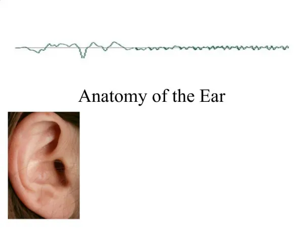
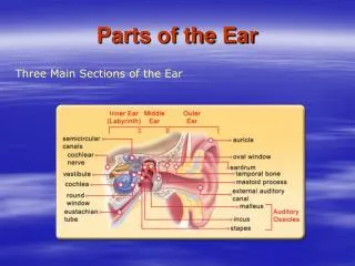
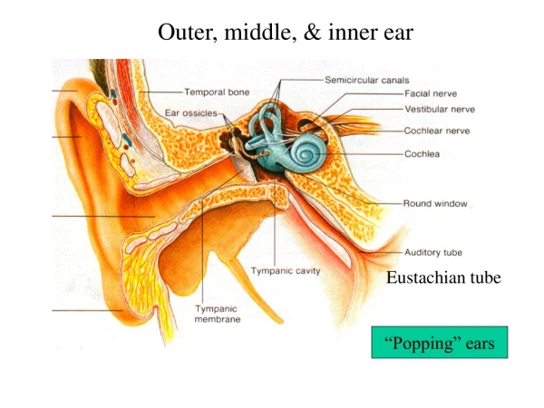
DEVELOPMENT OF EAR Ear is composed of three anatomical parts • External ear (Collection of sound waves) auricle, external auditory meatus, external layer of tympanic membrane • Middle ear (conduction of sound waves) 3 auditory ossicles, tympanic cavity, auditory tube • Internal ear (hearing & balance) vestibulocochlear organ which converts waves into nerve impulses & register changes in equilibrium
DEVELOPMENT OF INTERNAL EAR • Otic placodes -- on each side of myelencephalon • Inductive influence – notochord, paraxial mesoderm • Otic pit, otic vesicle, loses connection from surface ectoderm • Two parts of otic vesicle---- memb. labyrinth • Ventral component---- saccule & cochlear duct • Dorsal component---- utricle, semicircular canals, & endolymphatic duct
SACCULE, COCHLEA, & ORGAN OF CORTI • In 6thweek saccule forms a tubular out pocketing --- cochlear duct completes 2.5 turns in 8th week • Connected with saccule through ductus reuniens • Surrounding mesenchyme differentiates into cartilage • In10th week vacuoles develop in cartilage & two perilymphatic spaces formed • Scala tympani & scala vestibuli • Vestibular & basilar membranes • Spiral ligament, modiolus • Cells in wall of cochlear duct differentiate into organ ofcorti; forms two ridges inner ridge---- spiral limbus--- tectorial membrane Outer ridge---- 1 row of inner hair cells 3-4 rows of outer hair cells • Statoacoustic ganglion ---- wall of otic vesicle & neural crest cells
UTRICLE & SEMICIRCULAR CANALS • Semicircular canals (SSC) appear as out pocketings of utricular part of otic vesicle • Central parts of these diverticula fuse & disappear • Peripheral unfused parts of diverticula become SSC
One end of each canal dilates to form crus ampullare while crus nonampullare, does not widen. Since two of latter type fuse, however, only five crura enter the utricle, 3 with an ampulla & 2 without • Cells in the ampulla forms a crest, crista ampullaris containing sensory cells for maintenance of equilibrium • Similarly specialized receptor areas macula develop in utricle & saccule • Sensory areas are stimulated by positional changes & impulses re transmitted via vestibular ganglion to brain through CN- VIII
DEVELOPMENT OF MIDDLE EAR • Endoderm of 1st pharyngeal pouch forms the tubotympanic recess which comes in contact with 1st pharyngeal cleft to form the external auditory meatus • Distal end is expanded -------Tympanic cavity • Proximal end is narrow ----- auditory tube
EAR OSSICLES • Malleus & incus---cartilage of 1st pharyngeal arch • Stapes-----cartilage of 2nd pharyngeal arch • Remain embedded in mesenchyme till 8th month when surrounding tissue dissolves
Cont… • The endodermal epithelial lining of tympanic cavity then extends along the newly developing space • When ossicles are entirely free of surrounding mesenchyme, the endodermal epithelium connects them in a mesentery like fashion to the wall of cavity • Tympanic cavity expands dorsally by vacuolization of surrounding tissue to form tympanic antrum
DEVELOPMENT OF EXTERNAL EAR • Begins at 3rd month • External auditory meatus --- 1st pharyngeal cleft • Ectodermal cells at the bottom of cleft proliferate to form a solid epithelial plate, the meatal plug • During 7th month the plug dissolves to form internal part of external auditory meatus • Tympanic membrane is the 1st pharyngeal membrane b/w cleft & pouch
AURICLE • Develops from 6 mesenchymal proliferations in 1st & 2nd pharyngeal arches surrounding the first cleft • These swellings are auricular hillocks 3 on each side of external meatus
Cont… • These swellings later fuse to form definite auricle • As fusion of auricular hillocks is complicated developmental abnormalities of the auricle are common • Lobule is the last part to develop • As mandible develops the auricle moves to their normal position at the sides of head
CONGENITAL ANOMALIES • Congenital deafness Mal development of internal ear or auditory ossicles or ear drum • External ear defects Due to chromosomal abnormalities