Download

1 / 39
610 likes | 1.35k Views
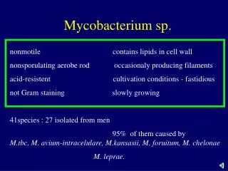
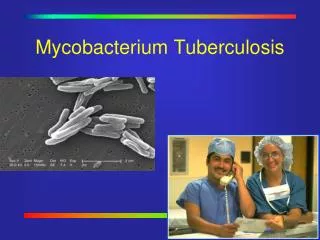
Mycobacterium. MYCOBACTERIUM. THIS GENUS IS COMPOSED OF: Strictly aerobic, acid-fast rods, does not Stain well (gram stain indeterminant), DNA has high g+c content, unique cell wall, Mycolic acid carbon chain length > c60 Relatively slow growth (two groups)

E N D
MYCOBACTERIUM THIS GENUS IS COMPOSED OF: Strictly aerobic, acid-fast rods, does not Stain well (gram stain indeterminant), DNA has high g+c content, unique cell wall, Mycolic acid carbon chain length > c60 Relatively slow growth (two groups) RAPID GROWERS(Visible colonies in <5 days) SLOW GROWERS(Visible colonies in > 5 days) TYPE SPECIES:Mycobacterium tuberculosis
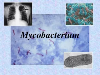
THE GENUS MYCOBACTERIUM CAN BE DIVIDED INTO FOUR BROAD GROUPS • THE TUBERCULOSIS COMPLEX • SLOW GROWING MYCOBACTERIA OTHER THAN TUBERCULOSIS (MOTT) • RAPIDLY GROWING MYCOBACTERIA • MYCOBACTERIUM LEPRAE
Acid Fastness Stain(Ziehl-Neelsen stain) • flood the slide with basic fuchsin (a red dye) in 5% phenol as a mordant. • heat gently for few minutes to melt the wax. • wash with 3% HCl in ethanol. • counter-stain with methylene blue. Mycobacterium stains red and other bacteria and the background are blue. The mycolic acid and its derivatives are responsible for the acid f
THE TUBERCULOSIS COMPLEX (Organisms that resemble M. tuberculosis; Causing a similar type of disease in humans) M. tuberculosis M. bovis
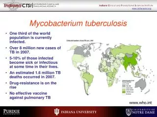
M. tuberculosisGeneral Features • It is a causative agent for human tuberculosis. • It grows very slow with a generation time of 12-15 hours. • On solid media the colonies are raised and rough with a wrinkled surface. • M. tuberculosis cells grow either as discrete rods or as aggregates. Virulent strains tend to grow as an aggregated long arrangement called serpentine cord. Cord factor is a derivative of mycolic acids, trehalose 6'-dimycolate.
Transmission • Through respiratory tract, alimentary tract, injured skin。 • TB in the lungs or throat can be infectious. This means that the bacteria can be spread to other people. TB in other parts of the body, such as the kidney or spine, is usually not infectious.
Who is at risk: Primary infection: children Secondary infection: age>25
Virulence factors No spore, no flagellum, no exotoxin,no endotoxin, no invasive enzyme • Capsule:polysaccharide;CR3;enzyme; protect • Lipid/Lipo arabinomannan • Heat-shock protein/Tuberculin protein: antigenicity, old tuberculin; associate with wax D can cause hypersensitivity and form tubercle
Lipid Lipid: closely related to virulence a. Phospholipid monocytes proliferate,cause tubercles b. Wax D adjuvent(not only to TB), delayed-type hypersensitivity c. Sulfatide硫酸脑苷脂 suppress phagosome combine with lysosome d. Cord factor (trehalose-6,6-dimycolate) destroy mitochondria, cause chronic granulomatosis, suppress WBC wandering
Pathogenesis primary infection 1) lung infection secondary infection 2) Out lung infection
Clinical syndromes a. fatigue, weakness, weight loss and fever b. pulmonary involvement: chronic cough,spit blood c. meningitis or urinary tract involvement d. bloodstream dissemination: miliary tuberculosis with lesions in many organs and a high mortality rate.
Primary Tuberculosis • The organisms are transmitted among human via aerosol. • TB bacilli lodge in the alveoli or lung alveolar ducts and most of bacilli are phagocytosed by alveolar macrophages. • Macrophages migrate to the hylar lymph node and generate T cell-mediated immune response. (can be monitored by tuberculin test)
Tuberculin Skin Test • Tuberculin is a mixture known as purified protein derivatives (PPD) from TB bacilli. • It is a test for delayed type hypersensitivity. Positive reaction, reddening and thickening (> 5mm) at the site of injection after 2-3 days, indicates cellular immunity to tubercle bacilli.
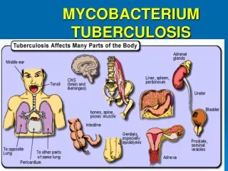
MYCOBACTERIUM TUBERCULOSIS Can infect (disseminate) and cause disease in many different body locations such as: Meninges Brain Bone Kidney Essentially any organ (lung primary target)
Steps in the development of tuberculosis Inhalation of bacteria Dead phagocytes, necrosis Bacteria reach lungs, enter macrophages M. tuberculosis Bacteria reproduce in macrophages Phagocytes, T cells, and B cells trying to kill bacteria Lesion begins to form (caseous necrosis) Activated macrophages Bacteria cease to grow; lesion calcifies Lesion liquefies Bacteria coughed up in sputum Immune suppression Spread to blood organs Reactivation Death
Immunity High rate of infection, but low morbidity. • Nonspecific immune AIDS, immunosuppressive agents, endocrine disease, etc.
Immunity-cellular Immunity First time: TB invade→proliferate on the spot →invade local lymph node Macrophage engulf TB →TH cell →IL-1 → TH proliferate →bloodstream Then TH meet TB again →MCF →macrophages congregate to focus →MAF →macrophages become more active →MIF →macrophages stay at the focus Then if it is successful granulomatosis forms,prevent TB diffusing;If it is not successful,macrophage can not kill TB, patients deteriorate.
Immunity • Cellular immunity 3-6 weeks, T cell VS macrophage 1. CD4+TH : INF-γ→macrophage→epithelioid cell granulomatosis 2. CD8 +TS : granule dependent, dissolve infected macrophage,kill TB 3. CD4- CD8 –t(γδ-T):Fas dependent, dissolve infected macrophage,but not kill TB, cause caseous focus in the center of granulomatosis; Acidity and lack of oxygen also make TB die.
Immunity • IV hypersensitivity Koch phenomenon; wax D+tuberculin protein; wax D →macrophage→epithelioid cell→tubercles→protect TB being phagocytized
Immunity • Humoral immunity A lot of Ab comes out, but meaningless TB active patient: immune complex more TB stable patient: immune complex less
Diagnosis The steps to diagnose TB infection and disease include: • A medical evaluation that includes history and risk assessment • The tuberculin skin test • A chest x-ray • A bacteriological examination
Diagnosis 1. Specimen: sputum, pus, CSF, urine, etc. 2. Microscopic examination: Ziehl-Neelsen stain 3. Concentration: 4%NaOH-3%HCL; 6% H2SO4 4. Culture: solid culture (2-4 weeks 37℃) ; liquid culture (1-2 weeks) 5. Animal inoculation: guinea pig 6. quick Diagnosis: PCR
Skin test PPD-C BCG-PPD >5mm + >15mm + + PPD-C>BCG-PPD infected
Mantoux method When the Mantoux skin test is performed, a needle is injected into the upper skin layer of the patient's arm. The arm is examined 48 to 72 hours after the tuberculin injection in order to evaluate the reaction on the patient's skin. Any swelling that can be felt around the site of the injection, also known as induration, is measured. The diagnosis of TB infection depends on the size of the measured induration and the patient's individual risk factors.
Prevention • BCG vaccination for new infants Freeze-drying vaccine rRNA vaccine eg:south India Chingleput’s failure of BCG • Find and cure patients
Treatment for Tuberculosis • Treated with a combination of multiple drugs for a long period of time: rifampin, isoniazid (INH), pyrazinamide, ethambutol, and streptomycin. • Emergence of multi-drug resistant M. tuberculosis strains.
Mycobacteria and AIDS • M. avium is much less virulent than M. tuberculosis • does not infect healthy people • infects AIDS patients • M. avium infects • when CD4 count greatly decreased • M. tuberculosis infection • infects healthy people • infects AIDS patients • earlier stage of disease • more systemic
Clinical features with AIDS • systemic disease (versus pulmonary) • greater in AIDS • lesions often lepromatous Antibiotic therapy • selected primarily for M. tuberculosis • if M. avium involved other antibiotics included
Mycobacterium avium-intracelluare complex • causes tb like disease in birds, opportunistic • pathogen in humans. Very prominent cause of • disease in aids patients has been decreased • following haart. Not easily transmitted. • (Runyon group III). Difficult to treat ( drug of • choice is rifabutin)
HANSEN’S DISEASE (Leprosy) caused by M. leprae • Hansen’s disease is a chronic, slowly progressive • granulomatous disease involving ectodermally derived • tissue such as the skin and peripheral nerves. The disease is usually limited to the cooler parts of the body such as the skin, nose and upper respiratory tract. It rarely affects internal organs such as the brain, liver, spleen, kidneys, and bones. • It has a specific predilection for peripheral nerves.
4 forms of Leprosy: Lepromatous Tuberculoid Borderline indeterminate