Download

1 / 61
660 likes | 1.53k Views
Mycobacterium. Important Human Pathogens. Mycobacterium tuberculosis Mycobacterium leprae (uncommon) Mycobacterium avium-intracellulaire Complex (MAC) or (M. avium). Mycolic acids. Lipid-Rich Cell Wall of Mycobacterium. CMN Group: Unusual cell wall lipids (mycolic acids,etc.).

E N D
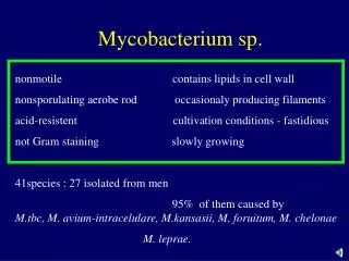
Important Human Pathogens Mycobacterium tuberculosis Mycobacterium leprae (uncommon) Mycobacterium avium-intracellulaire Complex (MAC) or (M. avium)
Mycolic acids Lipid-Rich Cell Wall of Mycobacterium CMN Group: Unusual cell wall lipids (mycolic acids,etc.) (Purified Protein Derivative)
Acid-Fast (Kinyoun) Stain of Mycobacterium NOTE: cord growth (serpentine arrangement)of virulent strains
Photochromogenic Mycobacterium kansasii on Middlebrook Agar • NOTE: Mycobacteria pathogenic for humans can be differentiated (Runyon Groups) by: • speed of growth (all are slower than most other pathogens) and by • production of chromogenic pigments (in light, in dark, or none)
Eight Week Growth of Mycobacterium tuberculosis on Lowenstein-Jensen Agar
Pathogenic Mycobacterium spp. BCG AIDS patients
Diagram of a Granuloma NOTE:ultimately a fibrin layer develops around granuloma (fibrosis), further “walling off” the lesion. Typical progression in pulmonary TB involves caseation, calcification and cavity formation.
Laboratory Diagnosis of Mycobacterial Disease Nucleic acid probes Nucleic acid sequencing
Differential Characteristics of Commonly Isolated Mycobacterium spp.
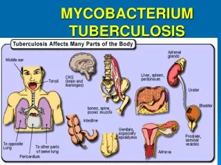
Mycobacterium tuberculosis Infections(cont.) Positive PPD + Chest X-Ray + MDR-TB a serious global health threat BCG(bacille Calmette-Guerin) = attenuated M. bovis
Typical Progression of Pulmonary Tuberculosis • Pneumonia • Granuloma formation with fibrosis • Caseous necrosis • Tissue becomes dry & amorphous (resembling cheese) • Mixture of protein & fat (assimilated very slowly) • Calcification • Ca++ salts deposited • Cavity formation • Center liquefies & empties into bronchi
PPD Tuberculosis Skin Test Criteria PPD = Purified Protein Derivative from M. tuberculosis
Tuberculoid vs. Lepromatous Leprosy Clinical Manifestations and Immunogenicity
Effect of Cell-Mediated Immunity on Leprosy Clinical Outcome
M. avium-intracellulaire Complex (MAC) Progression vs. CD4 Count in AIDS Patients
Mycobacterium avium-intracellulaire in Tissue Specimens High Magnification Low Magnification
REVIEW of Mycobacterium
Important Human Pathogens Mycobacterium tuberculosis Mycobacterium leprae (uncommon) Mycobacterium avium-intracellulaire Complex (MAC) or (M. avium) REVIEW
Mycolic acids Lipid-Rich Cell Wall of Mycobacterium CMN Group: Unusual cell wall lipids (mycolic acids,etc.) (Purified Protein Derivative) REVIEW
Pathogenic Mycobacterium spp. BCG AIDS patients REVIEW
Diagram of a Granuloma NOTE:ultimately a fibrin layer develops around granuloma (fibrosis), further “walling off” the lesion. Typical progression in pulmonary TB involves caseation, calcification and cavity formation. REVIEW
Mycobacterium tuberculosis Infections(cont.) Positive PPD + Chest X-Ray + MDR-TB a serious global health threat BCG(bacille Calmette-Guerin) = attenuated M. bovis REVIEW
Typical Progression of Pulmonary Tuberculosis • Pneumonia • Granuloma formation with fibrosis • Caseous necrosis • Tissue becomes dry & amorphous (resembling cheese) • Mixture of protein & fat (assimilated very slowly) • Calcification • Ca++ salts deposited • Cavity formation • Center liquefies & empties into bronchi REVIEW