Download
1 / 38
1.33k likes | 2.79k Views
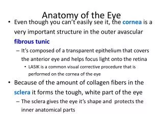
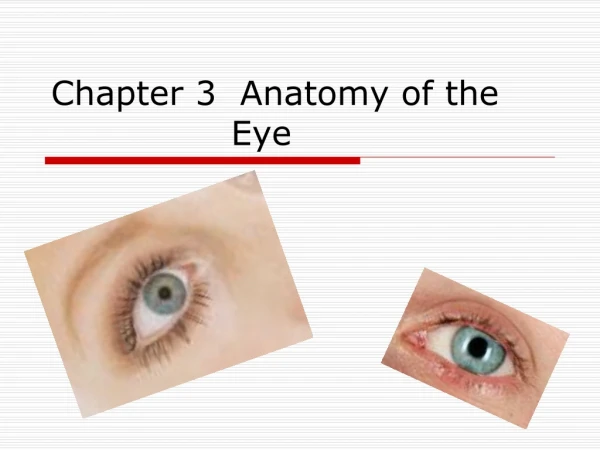
Anatomy of the Eye. The retina consists of two types of photoreceptor cells, rods and cones Rods are abundant in the periphery of the retina whereas cones are found more frequently in the central areas. Anatomy of the Eye.

E N D
Anatomy of the Eye • The retina consists of two types of photoreceptor cells, rods and cones • Rods are abundant in the periphery of the retina whereas cones are found more frequently in the central areas
Anatomy of the Eye • Each eye contains ≈ 120 million rod-shaped photoreceptors that are adapted for a low light threshold (high sensitivity) - they produce low resolution, black and white images • a loss of rods with age makes it difficult to drive at night
Anatomy of the Eye • Cone-shaped photoreceptors function in bright light to produce high resolution color images • They exists in three varieties, corresponding to the type of pigment they contain: red, green or blue • The photopigments are concentrated in the outer segment of the receptor, while the inner segment contains the nucleus and organelles
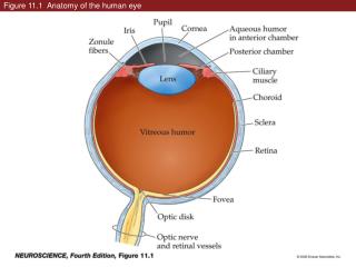
Eye Cavities and Chambers • The lens is an avascular refractory structure situated posterior to the pupil and iris. It consists of a capsule with crystallin proteins arranged in layers, and like the cornea, the lens is transparent • It attaches to the ciliary muscle of the ciliary body by suspensory ligaments that fine tune the focusing of light on the retina
Eye Cavities and Chambers • The lens divides the eyeball into two cavities: An anterior cavity anterior to the lens, and a posterior cavity (vitreous chamber) behind the lens • The anterior cavity is further divided at the level of the iris into anterior and posterior chambers (both filled with aqueous humor)
Eye Cavities and Chambers • The much larger posterior cavity of the eyeball (vitreous chamber) lies between the lens and the retina • Within the vitreous chamber is the vitreous body, a transparent jellylike substance that holds the retina flush against the choroid, giving the retina an even surface for the reception of clear images • occasionally, collections of debris called vitreal floaters cast shadows on the retina and create a spot in our field of vision (they are usually harmless and do not require treatment)
Eye Cavities and Chambers • This cow eye dissection shows an eye bisected into anterior and posterior sections along its coronal axis. The anterior structures of the iris and pupil are seen in the bottom half; the posterior retina, choroid, and optic disc are seen in the top half
Aqueous Humor • The eye requires a constant bath in a nourishing fluid to deliver enough O2 to support the avascular lens and cornea • It also needs fluid to help “inflate” the walls of the eyeball (maintain a constant intraocular pressure – IOP) and support the vitreous body • this need is accomplished through the production of aqueous humor, which flows through the anterior cavity of the eye and is replaced every 90 minutes
Aqueous Humor • Aqueous humor is produced at the ciliary body and flows first through the posterior chamber (of the anterior cavity of the eye) • Traveling along the posterior surface of the iris it passes through the pupil to enter the anterior chamber • It proceeds along the anterior surface of the iris until it is reabsorbed into the scleral venous sinus (canal of Schlemm) and returned to the venous system
Aqueous Humor • Any sort of blockage to aqueous humor flow, or overproduction at the ciliary body may result in an increase of pressure inside the eye – a condition called glaucoma • If not treated, glaucoma can lead to a degeneration of eye function
Retinal Detachment • The vitreous body (humor) also contributes to maintain proper intraocular pressure as it holds the retina against the choroid. The vitreous humor, however, is only formed during embryological development and is not replaced. As we age, shrinkage of the vitreous body may lead to a detachment of the retina from the choroid • A retinal detachment is considered a medical emergency and needs immediate repair before vision loss becomes permanent
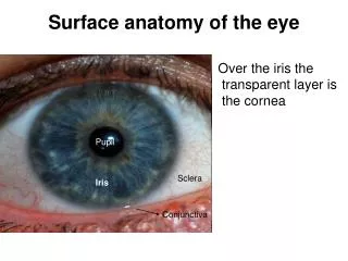
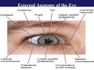
The Pupillary Response • The pupil is an opening in the center of the iris. It is composed of a radial muscle that “radiates” away from the center, and a circular muscle that is in the center • Contraction of the inner circular muscle fibers cause the pupil to constrict while contraction of the radial fibers cause it to dilate
Refraction and Image • Normal image formation depends on refraction of light waves, accommodation of the lens, constriction of the pupil, and convergence of the two eyes • Refraction is the process of bending light rays. Both the cornea and the lens refract light rays, and both must be functioning in order to properly focus light onto the right spot on the retina to produce clear vision
Refraction and Image • Since the cornea has a fixed shape, its “focal length” is also fixed; and its ability to refract light is likewise fixed • In order to focus light that has already been bent by the cornea the lens must change shape – the amount depending on the type of light rays we are trying to “see”
Refraction and Image • An increase in the curvature of the lens for near vision is called accommodation • The near point of vision is the minimum distance from the eye that an object can be clearly focused - about 4 in (a distance that increases with age due to a loss of elasticity in the lens)
Refraction and Image • Convergence is the inward movement of the eyes so that both are directed at the object being viewed - becoming a little cross-eyed when viewing things close up • The nearer the object, the greater the degree of convergence needed to maintain binocular vision • the coordinated action of the extrinsic eye muscles brings about convergence. • Convergence helps us maintain our binocular vision and see in three dimensions
Refraction and Image • With nearsightedness (myopia), only close objects can be seen clearly: Light rays coming in from distant objects are naturally focused in front of the retina and appear blurry • Correction involves the use of a concave (negative) lens • With farsightedness (hyperopia), only distant objects can be seen clearly: Light rays coming in from nearer objects are naturally focused behind the retina • Correction involves the use of a convex (positive) lens
Refraction and Image • Abnormal refractive capabilities of the eye are the result of a misshapen eyeball (usually too long or too short), or because the lens becomes stiff (usually with age). Corrections are accomplished using either a positive (convex) or negative (concave) lens (eyeglasses, contacts, or lens replacements)
Visual Transduction • Once light waves have been successfully focused on the retina, the information “stored” in that electromagnetic energy must be changed by photopigments in the photoreceptors into signals our brain can interpret - a process called visual transduction • The single type of photopigment in rods is rhodopsin, whereas there are 3 different cone photopigments • Color vision results from different colors of light selectively activating the different cone photopigments
Visual Transduction • The first step in visual transduction is absorption of light by a photopigment, a colored protein that undergoes structural changes when it absorbs light in the outer segment of a photoreceptor • Light absorption initiates a series of events that lead to the production of a receptor potential (number 4 in the diagram)
Visual Transduction • All photopigments associated with vision contain two parts: a glycoprotein known as opsinand a derivative of vitamin A called retinal • Although there are 4 different opsins, retinal is the light-absorbing part of all visual photopigments • To simplify the process we can say that there is a cyclical bleaching and regeneration of photopigment • Bleaching is a term describing a conformational change in the retinal molecule in response to light
Visual Transduction • In darkness, retinal has a bent shape called cis-retinal • Absorption of a photon of light causes it to straighten into the trans-retinal form in a process called isomerization • Trans-retinal completely separates from the opsin; since the final products look colorless, this part of the cycle is called bleaching of photopigment • An enzyme converts trans-retinal→ cis-retinal • The cis-retinal regenerates the photopigment
Bleaching and regeneration of photopigments are summarized here
Visual Transduction • In daylight, regeneration of rhodopsin cannot keep up with the bleaching process, so rods contribute little to daylight vision. In contrast, cone photopigments regenerate rapidly enough that some of the cis form is always present, even in very bright light • As a consequence, light adaptation (from dark conditions light conditions) happens in seconds; dark adaptation (from light dark) takes minutes to occur (up to 40 minutes to fully adapt)
Visual Transduction • Most forms of color blindness, an inherited inability to distinguish between certain colors, result from the absence or deficiency of one of the three types of cones • Most common type is red-green color blindness in which red cones or green cones are missing • Prolonged vitamin A deficiency and the resulting below-normal amount of rhodopsin may cause night blindness or nyctalopia, an inability to see well at low light levels
The Visual Pathway • The graded potentials generated by the photoreceptors undergo considerable processing at synapses among the various types of neurons in the retina (horizontal cells, bipolar cells, and amacrine cells)- certain features of visual input are enhanced while others are discarded • Overall, convergence pre- dominates as 126 million photo-receptors impinge on only 1 million ganglion cells
The Visual Pathway • The axons of retinal ganglion cells provide output that travels back “towards the light”, exiting the eyeball as the optic nerve,whichemerges from the vitreous surface of the retina • The axons then pass through a crossover point called the optic chiasm
The Visual Pathway • Some axons cross to the opposite side, while others remain uncrossed. Once through the optic chiasm the axons enter the brain matter as the optic tracts (most terminate in thalamus) • Here they synapse with neurons that project to the 1o visual cortex in the occipital lobes
Visual field of left eye Visual field of left eye Visual field of left eye Visual field of left eye Visual field of left eye Visual field of left eye Visual field of right eye Visual field of right eye Visual field of right eye Visual field of right eye Visual field of right eye Visual field of right eye Temporal half Temporal half Temporal half Temporal half Temporal half Temporal half Nasal half Nasal half Nasal half Nasal half Nasal half Nasal half Nasal half Nasal half Nasal half Nasal half Nasal half Nasal half Temporal half Temporal half Temporal half Temporal half Temporal half Temporal half Left eye Left eye Left eye Left eye Left eye Left eye Right eye Right eye Right eye Right eye Right eye Right eye Nasal retina Nasal retina Nasal retina Nasal retina Nasal retina Nasal retina Temporal retina Temporal retina Temporal retina Temporal retina Temporal retina Temporal retina Nasal retina Nasal retina Nasal retina Nasal retina Nasal retina Nasal retina Temporal retina Temporal retina Temporal retina Temporal retina Temporal retina Temporal retina 1 1 1 1 1 1 3 3 3 3 1 1 1 1 1 1 3 3 3 3 4 4 4 4 4 4 Optic tract Optic tract Optic tract 2 2 2 2 2 2 2 2 2 2 Midbrain Midbrain Midbrain Midbrain Midbrain Midbrain Midbrain Midbrain Midbrain Midbrain Midbrain Midbrain 5 5 5 5 Lateral geniculate nucleus of the thalamus Lateral geniculate nucleus of the thalamus Lateral geniculate nucleus of the thalamus Lateral geniculate nucleus of the thalamus Lateral geniculate nucleus of the thalamus Lateral geniculate nucleus of the thalamus 6 Optic radiations Optic radiations Optic radiations Optic radiations Optic radiations Optic radiations Optic radiations Optic radiations Optic radiations Optic radiations Optic radiations Optic radiations 6 Primary visual area of cerebral cortex (area 17) in occipital lobe Primary visual area of cerebral cortex (area 17) in occipital lobe Primary visual area of cerebral cortex (area 17) in occipital lobe Primary visual area of cerebral cortex (area 17) in occipital lobe Primary visual area of cerebral cortex (area 17) in occipital lobe Primary visual area of cerebral cortex (area 17) in occipital lobe Left eye and its pathways Left eye and its pathways Left eye and its pathways Left eye and its pathways Left eye and its pathways Left eye and its pathways Right eye and its pathways Right eye and its pathways Right eye and its pathways Right eye and its pathways Right eye and its pathways Right eye and its pathways
The Ear • Audition, the process of hearing, is accomplished by the organs of the ear. The ear is an engineering marvel because its sensory receptors can transduce sound vibrations with amplitudes as small as the diameter of an atom of gold into electrical signals 1000 times faster than the eye can respond to light • The ear also contains receptors for equilibrium
The Ear • The ear has 3 principle regions • The external ear, which uses air to collect and channel sound waves • The middle ear, which uses a bony system to amplify sound vibrations • The internal ear, which generates action potentials to transmit sound and balance information to the brain
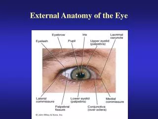
The External Ear • The anatomy of the external ear includes • The auricle (pinna), a flap of elastic cartilage covered by skin and containing ceruminous glands • A curved 1” long external auditory canal situated in the temporal bone leading from the meatus to the tympanic membrane (TM – or ear drum) which separates the outer ear from the cavity of the middle ear
The Middle Ear • The middle ear is an air-filled cavity in the temporal bone. It is lined with epithelium and contains 3 auditory ossicles (bones) • The stapes (stirrup) • The incus (anvil) • The handle of the malleus (hammer) attaches to the TM
The Middle Ear • Two small skeletal muscles (the tensor tympani and stapedius) attach to the ossicle and dampen vibrations to prevent damage from sudden, loud sounds
The Middle Ear • The Eustachian (auditory) tube connects the middle ear with the nasopharynx (upper portion of the throat) • It consists of bone and hyaline cartilage and is normally passively collapsed. It opens to equalize pressures on each side of the TM (allowing it to vibrate freely)
The Inner Ear • The internal ear (inner ear) is also called the labyrinth because of its complicated series of canals • Structurally, it consists of two main divisions: an outerbony labyrinththat encloses an inner membranous labyrinth • the bony labyrinth is sculpted out of the petrous part of the temporal bone, and divided into three areas: (1) the semicircular canals, (2) the vestibule, and (3) the cochlea