Download

1 / 55
590 likes | 699 Views
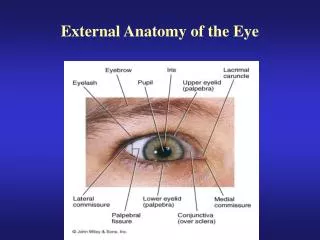
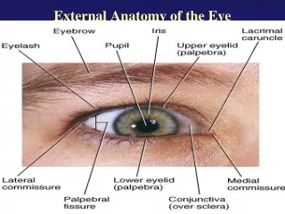
LEARN THIS!. EYE ANATOMY TUTORIAL. 1. Name the “third” eyelid (blue arrows). plica semilunaris. 2. Name the black area. cornea. 3. What are these “holes” called? (blue arrows)?. lacrimal tear ducts.

E N D
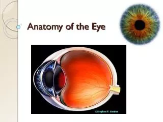
LEARN THIS! EYE ANATOMY TUTORIAL
1. Name the “third” eyelid (blue arrows). plica semilunaris
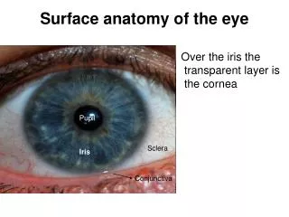
2. Name the black area. cornea
3. What are these “holes” called? (blue arrows)? lacrimal tear ducts
4. Name the structure represented by the “white stripes” embedded in the eyelids (blue arrows). Tarsal glands =Meibomian glands
5. In this animal the third eyelid is called a nictitating membrane. What structure in our eye may be evolutionarily derived from this structure?. plica semilunaris
6. What lines these three areas (blue arrows)? conjunctiva
7. Name this process. • Changing of lens shape to view objects Ciliary muscle fibers contracted Suspensory ligaments relaxed Lens thick accommodation (a) Ciliary muscle fibers relaxed Suspensory ligaments taut Lens thin (b)
8. Which eye demonstrates distant vision. eye 2 • Changing of lens shape to view objects Ciliary muscle fibers contracted eye 1 Suspensory ligaments relaxed Lens thick (a) eye 2 Ciliary muscle fibers relaxed Suspensory ligaments taut Lens thin (b)
9. Name this pitted area of the eye where your best vision occurs (blue arrows)? Fovea centralis
10. What are the structures shown by the blue arrows? ciliary body
11. Name this structure found in the back of the retina Macula lutea
13. Name the fluid in this part of the eye (blue arrow). Vitreous humor
14. Name this structure (blue circle). Optic nerve
15. Name the black area. choroid
Aqueous humor 16. Name the fluid shown circulating by the black arrows.
17. Name the circled part Optic disk
18. Name this area shown by blue arrow? Anterior chamber aqueous humor
19. Name this gelatinous part. Vitreous humor
20. What is the opening into the eye called (blue arrow)? pupil
21. What is the colored part of the eye called (blue arrow)? iris
22. What are the “string-like” structures shown by blue arrows? Suspensory ligaments
Name these cells shown by blue arrows? 23. Pigment cells of choroid
B 24. Name these muscles in order of the letters (no spaces). A C A =Lateral rectus B = Superior rectus C = Medial rectus D = Inferior rectus D Right eye
25. Name the fluid found in this area shown by blue arrow? Aqueous humor
26. Name this part. lens
Canal of Schlemm 30. Name this structure (yellow arrow)that drains the fluid shown circulating with the black arrows.
32. Name the shiny area. Tapedum lucidum
33. Name these muscles A and B in numerical order no spaces. A A = Superior oblique B = Inferior oblique B Right eye
35. Name this brownish structure. Extrinsic muscles
36. Name this entire structure. Ciliary body
37. Name this muscle. Levator palpebrae superioris
Ciliary body 38. Name this structure (yellow arrow)that produces the fluid shown circulating with the black arrows.
Suspensory ligaments 39. Name these string like structures.
20. Name this lining shown by green dotted line. conjunctiva
Tarsal glands 21. Name these structures that secrete oil.
22. Name this structures. Lacrimal gland
A. Plica semilunaris B. Lacrimal caruncle C. Lacrima papilla 23. Name these structures in order of the letters – no spaces. A B C
24. Name this opening. pupil
25. Name this part of the eye area. Optic nerve