Download
1 / 73
810 likes | 1.08k Views
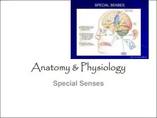
BASIC ANATOMY & PHYSIOLOGY OF THE EYE. Essam Osman , M.D. Professor & Consultant ophthalmologist Glaucoma unit. ANATOMY. EMBRYOLOGY OF THE EYE. This highly specialized sensory organ is derived from neural ectoderm, mesoderm and surface ectoderm.

E N D
BASIC ANATOMY & PHYSIOLOGY OF THE EYE EssamOsman, M.D. Professor & Consultant ophthalmologist Glaucoma unit
EMBRYOLOGY OF THEEYE • This highly specialized sensory organ is derived from neural ectoderm, mesoderm and surface ectoderm. • The eye is essentially an outgrowth from the brain (neural ectoderm). • Started as Optic vesicle connected to the forebrain by Optic stalk.
DEVELOPMENT OF THE EYE AFTER BIRTH • At birth, the eye is relatively large in relation to the rest of the body. • The eye reaches full size by the age of 8-13 years. • The lens continues to enlarge throughout the life. • The iris has a bluish color due to little or no pigment on the anterior surface. • During early infant life, the cornea & sclera can be stretched by raised IOP → enlargement of the eye.
THE ORBIT • As a socket, contains & protect the eye. • The weakest parts are the floor & the medial wall. • Seven bones contribute the bony orbit. • Surrounded by nasal sinuses. • Important openings are: • Optic foramen. • Superior orbital fissure. • Inferior orbital fissure.
THE EXTRAOCULAR MUSCLES • Four recti & two oblique muscles. • All are supplied by Oculomotor n. except superior oblique (Trochlear n.) & lateral rectus (Abducent n.).
Innervation & action of eye muscles: elevation intorsion extorsion abduction (lateral rotation) adduction (medial rotation) midline depression WHK CUHK 98
LR (6 n.) SO (4 n.) Innervation & action of eye muscles: IO SR (3 n.) midline MR IR
Hence for clinical test: (Field of Action) • Direction to look • SODown and in • IO Up and in • SR Up +/- out • IR Down +/- out
Globe Anatomy Or limbus
CONJUNCTIVA • Three parts: • Bulbar conjunctiva. • Palpebral conjunctiva. • Forniceal conjunctiva. • The stroma (no adenoid tissues until 3 months after birth). • Follicles & Papillae. • Injection and chemosis.
THE EYE (GLOBE) • Two spheres with different radii: - Cornea, window of the eye. - Sclera, opaque shell. *** The eye measures approximately 21-24 mm in all its main diameters.
The coats of the eye *** Three layers: • The outer: inelastic coat, transparent cornea and opaque sclera. • The middle, vascular coat, The Uvea: choroid, ciliary body and iris. • The inner: The Retina, extends forwards to within 6 mm of the limbus.
Cornea It has 5 layers. 500 -530 micron in thickness. Transparent Avascular Regularly arranged collagen fibers. Layers of cornea 1-epthelium 2-BM 3-stroma 4-DM 5-endothelium
The Chambers of The Eye ***Three optically clear spaces: • The anterior chamber, in front of the iris • The posterior chamber, immediately behind the iris. These two chambers which communicate through the pupil are filled with clear aqueous humour. • The vitreous cavity: filled by gel-like structure, The Vitreous. • ANTERIOR AND POSTERIOR SEGMENT
The Lens • The crystalline lens is the only structure continuously growing throughout the life. • Changeable refractive media. • Capsule, epithelium and lens fibers. • Congenital anomalies and effect of systemic diseases. • Cataract.
Retina and Vitreous • Vitreous attachment. • Optic nerve head, macula, fovea, retinal background, Ora serrata, and retinal vasculature. • Effect of systemic diseases. • Retinal detachment.
Optic Nerve contains around 1.2 million nerve fibers, which are axons of the retinal ganglion cells. 1 mm in the globe. 25 mm in the orbit. 9 mm in the optic canal. 16 mm in the cranial space Partial decussation occurs and about 53% of the fibers cross to form the optic tracts.
The Visual Pathway • Visual Pathway: Three neurons 1. Bipolar cell, lies within the retina. 2. Ganglion cell, synapse in lateral geniculate body. 3. Third neuron terminates in visual cortex.