Download

1 / 19
230 likes | 654 Views
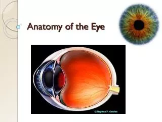
Eye Anatomy Notes. 1. Record this color key at the top of your paper : TUNICS- Layers of tissue forming the walls of the eye HUMORS- Fluids of the eye OPTICAL COMPONENTS- transparent elements that emit light rays and focus them on the retina NEURAL COMPONENTS Read the hint

E N D
Eye Anatomy Notes 1. Record this color key at the top of your paper: TUNICS- Layers of tissue forming the walls of the eye HUMORS- Fluids of the eye OPTICAL COMPONENTS- transparent elements that emit light rays and focus them on the retina NEURAL COMPONENTS Read the hint Use the word bank to identify the structure Record all information on your diagram
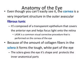
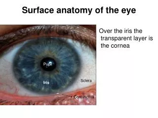
Hint: Transparent outer layer of the eye retina cornea fovea choroid coat Suspensory ligaments iris pupil sclera 1
Hint: Controls the amount of light entering the eye retina cornea fovea choroid coat Suspensory ligaments iris pupil sclera 2
Hint: Circular opening for light retina cornea fovea choroid coat Suspensory ligaments iris pupil sclera 3
Hint: Helps change the shape of the lens retina cornea fovea choroid coat Suspensory ligaments iris pupil sclera 4
Hint: Contains the photoreceptors retina cornea fovea choroid coat Suspensory ligaments iris pupil sclera 5
Hint: Contains blood vessels for nourishment and melanocytes to absorb excess light retina cornea fovea choroid coat Suspensory ligaments iris pupil sclera 6
Hint: Forms the white of eye, Protects eye, Provides attachment for muscles retina cornea fovea choroid coat Suspensory ligaments iris pupil sclera 7
Hint: Region of retina that produces the sharpest vision retina cornea fovea choroid coat Suspensory ligaments iris pupil sclera 8
Hint: Watery,Helps nourish space behind cornea Aqueous humor Vitreous humor 9 Hint: Jelly-like, Maintains the shape of the eyeball 10
Hint: Changes shape to focus light on the retina 11 Lens These structures also help focus light on the retina: Cornea Aqueous humor Vitreous humor
Cataracts appears as a cloudy lens. Click on the picture to view a video on cataract surgery. It’s Interesting!
Hint: Area where nerve fibers leave the retina and join the optic nerve Blind spot Optic nerve 13 12 Hint: Transmits messages to occipital lobe
The retina is also a neural component because it contains the photoreceptors: rods and cones. Rods sense black and white Cones sense color
Glaucoma: Damage to the optic nerve due to too much pressure. Click on the picture to watch the video.
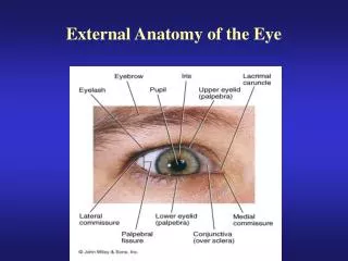
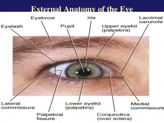
Accessory Organs of the Eye • Eyelid - protection • 2. Lacrimal apparatus • secretes tears which moistens the eye • tears contain enzymes that act as antibacterial agents • ducts drain the tears into the nasal cavity
Extrinsic muscles- move the eyeUse your knowledge of directional terms to determine the location of each extrinsic eye muscle. Superior rectus Superior oblique Inferior rectus Inferior oblique Lateral rectus Medial rectus
Think about what would happen if a particular muscle contracted. Match the action to the correct muscle. Abduction Adduction Depression Elevation Extorsion (rotates the top of the eye away from the nose) Intorsion (rotates the top of the eye toward the nose)
Conjunctivitis: Infection of the mucous membranes lining the eyelid. When you finish (and if you don’t have conjuctivitis) ask your teacher for homework. Homework is due next class.