Download
1 / 8
80 likes | 274 Views
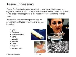
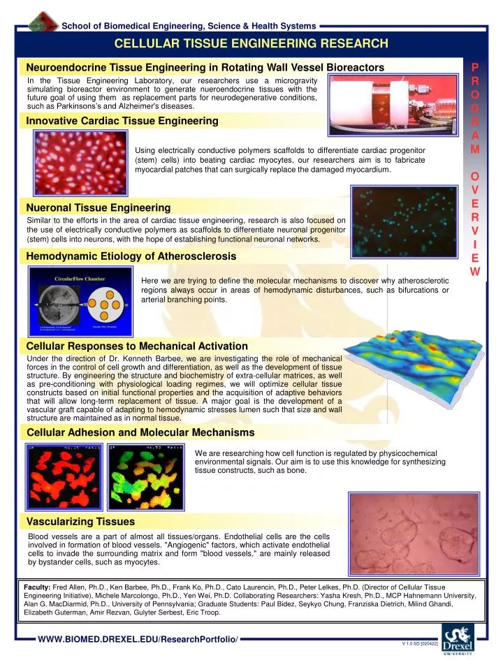
CELLULAR TISSUE ENGINEERING RESEARCH. Neuroendocrine Tissue Engineering in Rotating Wall Vessel Bioreactors. P R O G R A M O V E R V I E W.

E N D
CELLULAR TISSUE ENGINEERING RESEARCH Neuroendocrine Tissue Engineering in Rotating Wall Vessel Bioreactors P R O G R A M O V E R V I E W • In the Tissue Engineering Laboratory, our researchers use a microgravity simulating bioreactor environment to generate nueroendocrine tissues with the future goal of using them as replacement parts for neurodegenerative conditions, such as Parkinsons’s and Alzheimer's diseases. Innovative Cardiac Tissue Engineering • Using electrically conductive polymers scaffolds to differentiate cardiac progenitor (stem) cells) into beating cardiac myocytes, our researchers aim is to fabricate myocardial patches that can surgically replace the damaged myocardium. Nueronal Tissue Engineering • Similar to the efforts in the area of cardiac tissue engineering, research is also focused on the use of electrically conductive polymers as scaffolds to differentiate neuronal progenitor (stem) cells into neurons, with the hope of establishing functional neuronal networks. Hemodynamic Etiology of Atherosclerosis • Here we are trying to define the molecular mechanisms to discover why atherosclerotic regions always occur in areas of hemodynamic disturbances, such as bifurcations or arterial branching points. Cellular Responses to Mechanical Activation Under the direction of Dr. Kenneth Barbee, we are investigating the role of mechanical forces in the control of cell growth and differentiation, as well as the development of tissue structure. By engineering the structure and biochemistry of extra-cellular matrices, as well as pre-conditioning with physiological loading regimes, we will optimize cellular tissue constructs based on initial functional properties and the acquisition of adaptive behaviors that will allow long-term replacement of tissue. A major goal is the development of a vascular graft capable of adapting to hemodynamic stresses lumen such that size and wall structure are maintained as in normal tissue. Cellular Adhesion and Molecular Mechanisms We are researching how cell function is regulated by physicochemical environmental signals. Our aim is to use this knowledge for synthesizing tissue constructs, such as bone. Vascularizing Tissues Blood vessels are a part of almost all tissues/organs. Endothelial cells are the cells involved in formation of blood vessels. "Angiogenic" factors, which activate endothelial cells to invade the surrounding matrix and form "blood vessels," are mainly released by bystander cells, such as myocytes. • Faculty:Fred Allen, Ph.D., Ken Barbee, Ph.D., Frank Ko, Ph.D., Cato Laurencin, Ph.D., Peter Lelkes, Ph.D. (Director of Cellular Tissue Engineering Initiative), Michele Marcolongo, Ph.D., Yen Wei, Ph.D.Collaborating Researchers: Yasha Kresh, Ph.D., MCP Hahnemann University, Alan G. MacDiarmid, Ph.D., University of Pennsylvania; Graduate Students: Paul Bidez, Seykyo Chung, Franziska Dietrich, Milind Ghandi, Elizabeth Guterman, Amir Rezvan, Gulyter Serbest, Eric Troop.
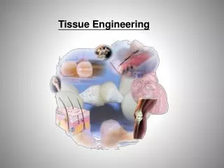
Functional Replacement Tissues Cellular / Molecular Biology: Development and Differentiation Stem Cells + Scaffolds / Nanotechnology Computer-Based Modeling Bioreactor Biotechnology (NASA) NEUROENDOCRINE TISSUE ENGINEERING IN ROTATING WALL VESSEL BIOREACTORS P R O J E C T O N E P A G E R • In the Tissue Engineering Laboratory, our researchers use a microgravity simulating bioreactor environment to generate nueroendocrine tissues with the future goal of using them as replacement parts for neurodegenerative conditions, such as Parkinsons’s and Alzheimer's diseases. • Faculty/Contact: P. Lelkes, Ph.D., Director of Cellular Tissue Engineering Laboratory, Drexel University. • E-mail: pilelkes@drexel.edu • Collaborating Researchers: F. Allen, Ph.D., Drexel University; K. Barbee, Ph.D., Drexel University. • Graduate Students: E. Troop, Drexel University; F. Dietrich, Drexel University. • Funding: NASA • Laboratories: Cellular Tissue Engineering Laboratory; MCP Hahnemann University.
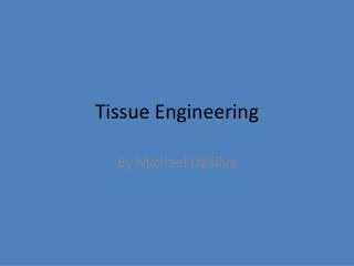
RAMEC on Electrically Conductive Polymer Thick film- Hoechst/Rhodamine • Thin film – • Phase Contrast Bis-Benzamide nuclear staining of H9C2 myoblast cells on 1M HCl doped polyaniline film. Rhodamine-phalloidin stained H9C2 myoblast cells on 1M HCl doped polyaniline film. INNOVATIVE CARDIAC TISSUE ENGINEERING • Using electrically conductive polymers as scaffolds to differentiate cardiac progenitor (stem) cells) into beating cardiac myocytes, our aim is to fabricate myocardial patches that can surgically replace the damaged myocardium. P R O J E C T O N E P A G E R • Cardiac Tissue: Hypothesis • Our working hypothesis states that tissue-specific differentiation and function of pluripotent stem cells into cardiac myocytes can be fine-tuned by the molecular structure/composition of 3-D scaffolds on the nano-scale. • Specifically, we hypothesize that 3-D scaffolds, composed of nanofibers made of, or doped with, electrically conductive polymers will, in conjunction with advanced RWV Bioreactor Biotechnology, facilitate the differentiation of cultured pluripotent mesenchymal stem cells into cardiac myocytes. Moreover, the application of controlled levels of electrical current will enhance/accelerate the formation of functional (beating) 3-D cardiac tissues. • Cardiac Tissue: Aims • Specific aim 1: Generate 3-D nanoscale scaffolds composed of electrically conductive polymers, alone or composite, and to optimize adhesion, growth, and differentiation of pluripotent stem cells and cardiac myocytes (isolated from adult or neonatal rats) on these scaffolds. • Specific aim 2: Compare tissue-specific differentiation into functional cardiac tissues by exposing the cells to electrical current in conventional cultures and in RWV Bioreactors. • Specific aim 3: Generate a functional vasculature within the tissue constructs. e-Pan We are currently testing new techniques to increase the biocompatibility of the electroactive PAN(e-PAN). A serial dedoping technique yields a substrate with improved cell adhesion and proliferation. As seen in figures 5 and 6, H9C2 myoblasts adhere and form confluent monolayers on the e-PAN. • Faculty/Contact: P. Lelkes, Ph.D., Director of Cellular Tissue Engineering Laboratory, Drexel University. • E-mail: pilelkes@drexel.edu • Collaborating Researchers: Y. Kresh, Ph.D., Drexel University; Y. Wei, Ph.D., Drexel University; F. Ko, Ph.D., Drexel University; • Alan MacDiarmid, Ph.D., University of Pennsylvania. • Graduate Students: P. Bidez, Drexel University; F. Dietrich, Drexel University. • Funding: NASA, Synergy Grant. • Laboratories: Cellular Tissue Engineering Laboratory; MCP Hahnemann University.
To study cell attachment on PANi and chloromethylated PANi films the wells containing various films made from commercially available PANi, chloromethylated PANi and PANi modified with tryptophan were seeded with PC12 cells. The wells were rinsed with PBS to remove cells that did not adhere to the films. The adherent cells were fixed and stained. To visualize the actin cytoskeleton we labeled actin filaments with rhodamine phalloidin, which specifically stains F-actin. At the same time cells were also stained with bis-benzamide stain, which is a nuclear stain. Upon staining cells were visualized using fluorescence microscopes. (a) (b) PC12 cells growing on the films, original magnification 100 X: (a) PC12 cells on unmodified PANi film, BBZ stain for cell nucleus. (b) PC12 cells on tryptophan modified PANi film, BBZ stain for cell nucleus. (c) PC12 cells on tryptophan modified PANi film, Rhodamin- Phalloidin stain for cytoskeletal F-actin. (c) NEURONAL TISSUE ENGINEERING P R O J E C T O N E P A G E R • To be successful, tissue engineering attempts to recapitulate some of the fundamental steps in organ development, such as electrical current-driven differentiation in the nervous system. Application of conducting polymers in biotechnology remains a challenge because of the supposedly poor biocompatibility of these materials. We hypothesize that the biocompatibility of electroactive polymers, such as polyaniline (PANI), can be improved by covalently linking bioactive peptides onto the surfaces of prefabricated conducting polymer films/fibers. Our long-term goal is to generate 3-D scaffolds from biocompatible conductive PANI that exhibit enhanced adhesion, proliferation, and differentiation of cells seeded onto them. • In this study, we demonstrated the feasibility of our approach by covalently linking a model amino acid, tryptophan, to the chloromethylated PANI backbone. The structure of the modified PANI was assessed using UV-VIS, IR, and NMR spectroscopy. The fluorescence properties of tryptophan allow for quantitative validation of its covalent attachment to the polymer. In assessing the biocompatibility of this modified conductive PANI, we are currently testing attachment, proliferation and differentiation of a cellular model for neuronal differentiation, PC-12 pheochromcytoma cells. In comparison to unmodified PANI, PC-12 cells seeded onto films prepared from the modified polymers exhibit enhanced attachment and proliferation, in addition to remaining responsive to nerve growth, factor-induced neuronal differentiation. This significantly improved PANI is now available for 3-D scaffold formation (e.g. by electrospinning of nanofibers) and for a variety of biomedical engineering applications, such as generating replacement tissues in neurodegenerative diseases, as well as spinal cord injury. • Faculty/Contact: P. Lelkes, Ph.D., Director of Cellular Tissue Engineering Laboratory, Drexel University. • E-mail: pilelkes@drexel.edu • Collaborating Researchers: Y. Kresh, Ph.D., Drexel University; Y. Wei, Ph.D., Drexel University; F. Ko, Ph.D., Drexel University; • Alan MacDiarmid, Ph.D., University of Pennsylvania. • Graduate Students: Shan Cheng, Paul Bidez, and Kolby Palouian (Undergraduate Student) - Drexel University. • Funding: NASA, US Army Research Office, Synergy Grant, and The Nanotechnology Institute. • Laboratories: Cellular Tissue Engineering Laboratory; MCP Hahnemann University.
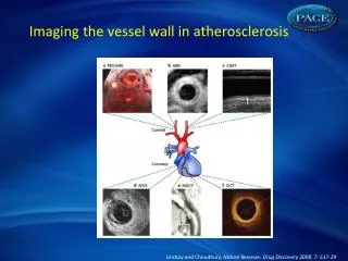
2 1 5 3 4 Shear Forces: 0.01-2.5 dynes/cm2 Pulsatile, Perturbed Flow, 100 ml/min HEMODYNAMIC ETIOLOGY OF ATHEROSCLEROSIS P R O J E C T O N E P A G E R Disturbed Hemodynamic Forces Predispose Endothelial Cells to Atherosclerotic Lesions In Vitro System Models Events at Sites of Atherosclerotic Predilection Atherosclerotic injury occurs at sites of hemodynamic disturbances (e.g., at branching points or bifurcations). Enhanced monocyte adhesion: First step towards developing atherosclerotic lesions. Model chamber for exposing cultured endothelial cells to disturbed flow. Altered gene expression: Analysis by cDNA micro-arrays. • Faculty/Contact: P. Lelkes, Ph.D., Director of Cellular Tissue Engineering Laboratory, Drexel University. • E-mail: pilelkes@drexel.edu • Collaborating Researchers: F. Allen, Ph.D., Drexel University; K. Barbee, Ph.D., Drexel University. • Graduate Students: P. Bidez, E. Troop, A. Rezvan - all from Drexel University. • Funding: Berlex Biosciences, Richmond, CA. • Laboratories: Cellular Tissue Engineering Laboratory; MCP Hahnemann University.
3-D surface topography of an endothelial monolayer aligned by flow in vitro. CELLULAR RESPONSES TO MECHANICAL ACTIVATION P R O J E C T O N E P A G E R The promise of tissue engineering lies in the prospect of replacing tissue that has become dysfunctional due to trauma or disease with new tissue capable of responding and adapting to environmental stimuli. Of primary importance in tissues that serve a mechanical or structural function is the ability to sense and respond to mechanical forces in order to adapt to the changing physical demands on the tissue. Previous in vivo and in vitro studies suggest that the structure and mechanical properties of blood vessel walls develop in response to the stress history of the tissue. The endothelium mediates vascular tone and structural remodeling in response to changes in blood flow, while the vascular smooth muscle (VSM) cells sense and respond to changes in stress within the vessel wall itself. These responses are essential to the maintenance of structural integrity and the regulation of blood flow. The central hypothesis of this research is that in normal development, structural relationships in vascular tissue are optimized for efficient sensing and transduction of the mechanical environment by the cells of the vessel wall. To engineer a tissue structure intended to acquire the property of adaptability present in normal tissue, we must first understand the salient features of the cells’ interaction with their surrounding structures that allow appropriate mechano-transduction to occur. The structure and biochemistry of engineered matrices, as well as pre-conditioning with physiological loading regimes, will be analyzed and optimized based on initial functional properties and the acquisition of adaptive behaviors that will allow long-term replacement of tissue. • Faculty/Contact: P. Lelkes, Ph.D., Director of Cellular Tissue Engineering Laboratory, Drexel University. • E-mail: pilelkes@drexel.edu • Collaborating Researchers: K. Barbee, Ph.D., Drexel University; F. Allen, Ph.D., Drexel University. • Graduate Students: A. Rezvan, Drexel University; E. Troop, Drexel University. • Funding: Berlex Biosciences, Richmond, CA. • Laboratories: Cellular Tissue Engineering Laboratory; MCP Hahnemann University.
Physical Factors : Fluid Flow and Biochemical Signaling Serum-free response Serum-supplemented response (2%) 1 min. after 1.5 min. after peak 20 sec. baseline flow flow after flow Intracellular calcium concentrating response to laminar fluid flow, with and without cell culture serum. CELLULAR ADHESION AND MOLECULAR MECHANISMS P R O J E C T O N E P A G E R We are researching how cell function is regulated by physicochemical environmental signals. Our aim is to use this knowledge for synthesizing tissue constructs, such as bone. • We use time-lapse and fluorescence • mcroscopy to study the biophysical • and biochemical function of cells, such • as: • Cell Adhesion • Cell Migration • Intracellular Signaling • Faculty/Contact: P. Lelkes, Ph.D., Director of Cellular Tissue Engineering Laboratory, Drexel University. • E-mail: pilelkes@drexel.edu • Collaborating Researchers: F. Allen, Ph.D., Drexel University. • Graduate Students: Amir Rezvan, Drexel University. • Funding: • Laboratories: Cellular Tissue Engineering Laboratory; MCP Hahnemann University.
Vascularizing Tissues : Blood vessels are a part of almost all tissues/organs. Endothelial cells are the cells involved in formation of blood vessels. "Angiogenic" factors, which activate endothelial cells to invade the surrounding matrix and form "blood vessels," are mainly released by bystander cells, such as myocytes. We are using cultured human microvascular endothelial cells (HMECs) to study mechanisms of neovascularization of ‘tissue constructs’ in vitro. The aim is to identify factors which cause HMECs to invade a three dimensional matrices made of fibrin, collagen or Matrigel and form "vascular networks." Ganglia-like structures formed by PC12 cells in coculture with HMEC cells after 3 days in fibrin gel. Cell-Cell Interactions : Every organ in the human body consists of several cell types which mutually influence each other. Our goal is to "engineer: functional cardiovascular tissues, We established cocultures of cells modeling the constituent cells of a heart, myocytes (H9C2), endothelial cells(HMEC) and nerve cells (PC12) by growing them in 3-dimensional fibrin gel matrices. We study phenotypic and genotypic changes in these cells and how they release specific factors, such as growth factors and proteolytic enzymes. The aim is to establish 3-D coculture conditions which will result in the correct activation of tissue specific signaling pathways and ultimately in functional tissue-like constructs for implantation or pharmacological studies. Tube-like structures formed by cardiac myoblasts in coculture with PC12 cells after 10 days in fibrin gel. VASCULARIZING TISSUES P R O J E C T O N E P A G E R • Faculty/Contact: P. Lelkes, Ph.D., Director of Cellular Tissue Engineering Laboratory, Drexel University. • E-mail: pilelkes@drexel.edu • Collaborating Researchers: Franziska Dietrich, Graduate Student, Drexel University. • Funding: • Laboratories: Cellular Tissue Engineering Laboratory; MCP Hahnemann University.