Download
1 / 34
350 likes | 385 Views
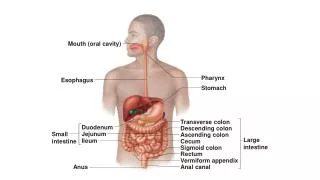
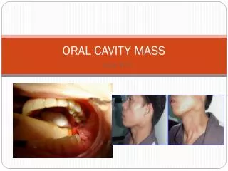
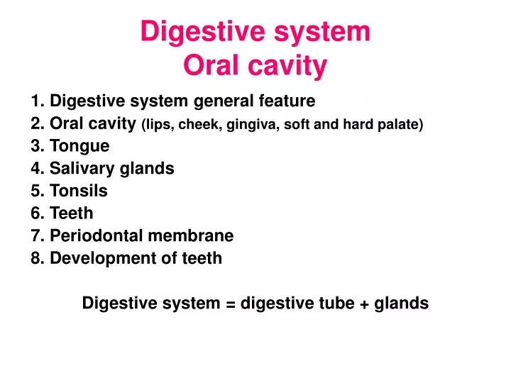
Digestive system Oral cavity. 1. Digestive system general feature 2. Oral cavity (lips, cheek, gingiva, soft and hard palate) 3. Tongue 4. Salivary glands 5. Tonsils 6. Teeth 7. Periodontal membrane 8. Development of teeth Digestive system = digestive tube + glands. Oral cavity.

E N D
Digestive systemOral cavity 1. Digestive system general feature 2. Oral cavity (lips, cheek, gingiva, soft and hard palate) 3. Tongue 4. Salivary glands 5. Tonsils 6. Teeth 7. Periodontal membrane 8. Development of teeth Digestive system = digestive tube + glands
Oral cavity Functions І. Main 1. Digestion (ingestion, mastication) 2. Moistening of food ІІ. Additional1. Articulation 2. Facial expression 3. Sensory reception 4. Respiratory І. Mucosa 1. Еpithelium – stratified squamous nonkeratinized epithelium (180-600 mkm) 2. Lamina propria – loose connective tissue 3. Muscularis mucosa-smooth muscular tissue ІІ. Submucosa - loose connective tissue (glands)
Lips • Hear-bearing skin: а) epitheliumstratified squamous keratinized (4 layers) • b) lamina propria(loose connective tissue with hair folliclessweat and sebaceous glands) 2. Transitional(vermilion):external (smooth) and internalzone 3. Mucosal а) epitheliumstratified squamous nonkeratinized b) lamina propria (papillae) • c) submucosa
Lip Red border of lip Papillae of connective tissue Blood vessel Musculus orbicularis oris Minor salivary gland Hair follicles Skin surface Mucosa surface Sweat gland
Tongue Functions 1. Mechanical 2. Receptory (general and taste) 3. Articulation Tunices І. Mucosa ІІ. Submucosa ІІІ. Muscular ІV. Aponeurosis Morphofunctional peculiarities of the lover surface of the tongue • Thin permeable epithelium • Well prominent blood supply • Salivary glands
Cheeks Cheek surfaces 1. External (skin) 2. Internal (mucosa) • І. Skin • ІІ. Muscle • ІІІ. Mucosa Mucosa zones 1. Upper(maxillary) 2. Lover (mandibulary) 3. Intermediate (no salivary glands,parotid glands ducts) 1. Mucosa • а) stratified squamous nonkeratinized epithelium (smear); • б) lamina propria (dense connective tissue, elastic fibers). 2. Submucosa(salivary glands). 3. Muscular tunic(skeletal muscle).Adipose body of Bisha.
Palate • Separates oral and nasal cavity • І. Hard palate • Zones1. Adipose • 2. Mucous • 3. Marginal • 4. Middle line(epithelial bodies) • ІІ. Soft palate • Oral surface(strutified squamous nonkeratinized epithelium) • Nasal surface(ciliated epithelium)
Tonsils • Lymph epithelial ring of Pirogov-Waldeier 7 tonsils : 2 tubular, 2 palatine, lingual, pharyngeal, laryngeal Functions 1. Protective 2. Immune І. Mucosa: а) epitheliumstratified squamous nonkeratinized infiltrated b) lamina propria – loose connective tissue with B lymphocytes) ІІ. Submucosa – septae, capsule ІІІ. Muscular tunicskeletal muscle
Tonsil Stratified squamous nonkeratinized epithelium Skeletal muscle Crypt Connective tissue septae Lymph nodules Crypt
Salivary glandsmajor (3pairs) + minor Functions 1. Exocine (saliva secretion) 2. Desinfection 3. Digestion 4. Articulation 2. Endocrine (parotin, VIP, glukagon, kallikrein, renin ...) 3. Excretion 4.Homeostatic Saliva Water -99% Minerals (Na, K, Cа, Fe,I…) Organic matters (enzymes, mucous) Cells (leucocytes, epithelial) MFU– Acinus= secretory portion+ intercalated duct+ striated duct
Myoepithelial cells Intercellular canaliculi Serocytes Myoepithelial cells Mixed acinus Serous acinus Serous demilunar Intercalated duct Intercalated ducts Striated duct Basement membrane Mucous acinus Mucocytes
Teeth • Functions • Structure • Deciduous and permanent teeth • Enamel • Dentin • Cementum • Pulp • Periodontal membrane • Development of teeth
Functions • Mastication • Articulation • Esthetic
Lip, tooth, gingiva red boarder enamel dentin pulp gingiva gingival fissure minor salivary glands periodontum skeletal muscle channel of tooth alveolar bone cementum lip furrow mandible tooth apex skin with hair muscle
Deciduous teeth (20) 1. Smaller 2. Crown lower and wider 3. Shorter roots Incisors central and lateral Cuspids (canines) Molars Eruption – 6 months- 2 years 12 years Permanent teeth (32) Incisors Cuspids Bicuspids (premolar)s Molars Eruption - 2012/2102 2012/2102 3212/2123 3212/2123
Enamel rods Enamel • Covers crown • 2,5 mm • Hardest tissue Chemical composition 1) 96-97 % – mineral salts (Са hydroxyapatites, fluorides, carbohydrates) 2) 3-4 % – organic substance (fibrillar matrix) MFU – hexagonal enamel rod (S-shaped enamel prism) Retzius, 1835 5-12 mln in tooth 4-8 мm
Enamel organ reticulum Basal terminal bar Mitochondria Rods are formed by ameloblasts Fur – microorganisms and + mineral and organic substances Pellicle - glycoproteins (II keratinous membrane) Cuticle – ameloblats remnants (I Nasmyth’s membrane) Nucleus RER Golgi complex Apical terminal bar Secretory granules Tome’s process Enamel
Enamel Enamel lamellae –thin leaflike structures (septae) Enamel bundles - near dentino- enamel junction Enamel tuft (spindle) Retzius incremental lines Hunter-Schreger bands Dentin
Dentin Chemical composition 1) 72 % – mineral salts (Са and Mg phosphates, carbohydrates, fluorides) 2) 28 % – organic substance (collagen fibers) Calcified dentin - dentinal tubules with Tomes’ fibers Mantle dentin (radial Korff’s fibers) Inner dentin (tangential Ebner fibers – imbrication lines) Contour lines of Owen Uncalcified dentin – predentin (odontoblasts in pulp)
Dentin cross section Dentin tubul Odontoblast process Peritubular dentin Dentin tubules Intertubular dentin Peritubular dentin – Neumans’ sheath (less fibers) Intertubular dentin – contains calcium crystals and globules (linear and globular calcification) Interglobular dentin – hypocalcified ground substance Tomes’ granular layer- a thin layer of dentin adjacent to cementum (in the root) Peritubular dentin Intertubular dentin
Dentin Predentin Odontoblasts Pulp Odontoblasts process Odontoblast nucleus
Dentin tubule Calcification zone Dentin Odontoblast procecss Odontoblast • Lies in the pulp • Produces dentin • Promotes regeneration Predentin Terminal bar RER GA RER Mitochondria Odontoblast nucleus
Primary dentin - regular Secondary dentin – forming pulpward of the demarcation line (on the pulpal surface) Transparent dentin – sclerotic (Ca salts are deposed in tubule and oblitarate it ) Dead tract(black)-empty tubules are filled with air Denticles – pulp stones 1. True (tubuli and cells) 1. Free (in pulp) 2. False (concentric calcified) 2. Adherent (attached) 3. Diffuse calcifications 3. Embedded (in dentin)
Cemento-enamel junction Cementum 1835 two Purkinje pupils • Cellular (cementocytes) • Acellular Chemical composition 1. 65 % – inorganic substances (Са(РО3)2, СаСО3) 2. 23 % – organic material (collagen fibers and ground substance) 3. 12 % – water Odontoblasts Root Apical foramen
Pulp – soft tissue Functions 1. Formative 2. Nutritive 3. Sensory 4. Protective In the crown In the root
Pulp layers • Peripheral – odontoblasts and immature collagen fibers 2. Intermediate – preodontoblasts and precollagen fibers • Acellular zone of Weil (cell-free layer in the crown of old teeth) 3. Central– loose connective tissue with blood vessels and nerves
Periodontal membrane (ligament) 1. Formative 2. Supportive 3. Sensory 4. Nutritive Epithelial rests of Malassez (Hertwig’s epithelial root sheath) Cementicles
Parodontum = gingiva + alveola + periodontum Circular ligament (10 ) Periodontum: 1) marginal (4) 2) proper (2)
Developmentalstages1. Dental laminaand bud2. Enamel organ(cap and bell stage)3. Histogenesis, organogenesis Low lip Low lip Deciduous tooth bud Tongue Dental Lamina DL DP Enamel organ Mesenchyme Dental papilla Maxilla A B Maxilla Dental lamina Reduced EO DL remnant Enamel organ Permanent tooth Enamel organ P Enamel Enamel Dentin Dentin Pulp Dental papilla Dental papilla P Dental sac C D Maxilla
Deciduous tooth Zone of eruption Permanent tooth Enamel Enamel Dentin Pulp Permanent tooth Enamel organ Dentin Pulp Periodontum Enamel Tooth eruption and replacement 1870 Hunter – root growth 1929,1936 Jasvoin – dental pupilla 1940 Kutz – bony tissue +pressure in pupilla Enamel Dentin Pulp Osteoclasts Periodontum Dentin Dental sac Pulp Dental sac