Download
1 / 46
480 likes | 723 Views
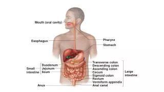
Oral Cavity II. Dr. Nabil Khouri MD, PhD. The Tongue. Mass of striated muscles covered with the mucous membrane Divided into right and left halves by a median septum Three parts: Oral (anterior ⅔) Pharyngeal (posterior ⅓) Root (base) Two surfaces: Dorsal Ventral. Functions.

E N D
Oral Cavity II Dr. Nabil Khouri MD, PhD
The Tongue • Mass of striated muscles covered with the mucous membrane • Divided into right and left halves by a median septum • Three parts: • Oral (anterior ⅔) • Pharyngeal (posterior ⅓) • Root (base) • Two surfaces: • Dorsal • Ventral
Functions • The tonge is the most important articulator for speech production. During speech, the tongue can make amazing range of movements • The primary function of the tongue is to provide a mechanism for taste. Taste buds are located on different areas of the tongue, but are generally found around the edges. • They are sensitive to four main tastes: Bitter, Sour, Salty & Sweet
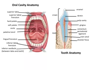
Dorsal Surface • Divided into anterior two third and posterior one third by a V-shaped sulcusterminalis. • The apex of the sulcus faces backward and is marked by a pit called the foramen cecum • Foramen cecum, an embryological remnant, marks the site of the upper end of the thyroglossal duct • Anterior two third: mucosa is rough, shows three types of papillae: • Filliform • Fungiform • Vallate • Posterior one third: No papillae but shows nodular surface because of underlying lymphatic nodules, the lingual tonsils
PAPILLAE • CIRCUMVALLATE PAPILLAE are arranged in a row parallel to and in front of sulcusterminalis • FUNGIFORM PAPILLAE are numerous at the tip and margin of the tongue. • FILLIFORM PAPILLAE are prevalent on the • dorsum of the tongue arranged in rows parallel to sulcus terminalis
Ventral Surface • Smooth (no papillae) • In the midline anteriorly, a mucosal fold, frenulum connects the tongue with the floor of the mouth • Lateral to frenulum, deep lingual vein can be seen through the mucosa • Lateral to lingual vein, a fold of mucosa forms the plica fimbriata
Muscles • The tongue is composed of two types of muscles: • Intrinsic • Extrinsic • The intrinsic M • Confined to tongue • No bony attachment • Consist of: • Longitudinal fibers • Transverse fibers • Vertical fibers • Function: Alter the shape of the tongue
INTRINSIC MUSCLES Muscles of the tongue All intrinsic muscles are supplied by Hypoglossal nerve
Extrinsic Muscles • Connect the tongue to the surrounding structures: the soft palate and the bones (mandible, hyoid bone, styloid process) • Include: • Palatoglossus • Genioglossus • Hyoglossus • Styloglossus • Function: Help in movements of the tongue
Movements • Protrusion: • Genioglossus on both sides acting together • Retraction: • Styloglossus and hyoglossus on both sides acting together • Depression: • Hyoglossus and genioglossus on both sides acting together • Elevation: • Styloglossus and palatoglossus on both sides acting together
Sensory Nerve Supply • Anterior ⅔: • General sensations: Lingual nerve • Special sensations : chorda tympani • Posterior⅓: • General & special sensations: glossopharyngeal nerve • Base: • General & special sensations: internal laryngeal nerve
Motor Nerve Supply • Intrinsic muscles: • Hypoglossal nerve • Extrinsic muscles: • All supplied by the hypoglossal nerve, except the palatoglossus • The palatoglossus supplied by the pharyngeal plexus
Blood Supply Dorsal lingual artery & vein Lingual artery & vein • Arteries: • Lingual artery • Tonsillar branch of facial artery • Ascending pharyngeal artery • Veins: • Lingual vein, ultimately drains into the internal jugular vein Deep lingual vein Hypoglossal nerve
Lymphatic Drainage • Tip: • Submental nodes bilaterally & then deep cervical nodes • Anterior two third: • Submandibular unilaterally & then deep cervical nodes • Posterior third: • Deep cervical nodes (jugulodigastric mainly)
Specialized mucosa • Covers the dorsum of the tongue. • Occupies 15% of the oral cavity. • Although it is masticatory mucosa by function but due to its high extensibility and lingual papillae, it is classified as • “SPECIALIZED MUCOSA”. • Lingual Papillae: These are the small nipple or hair–like structures on the upper surface of the tongue that give the tongue its characteristic rough texture. Four types of papillae are found on dorsum of the tongue: 1. Fungiform papillae 2. Filiform papillae 3. Foliate papillae 4. Circumvallate papillae
Fungiform Papillae: fungus-like appearance present on tip and sides of tongue scattered between filiform papillae smooth, rounded structures covered by non-keratinized epithelium Appear red due to highly vascular CT Taste buds are present on the superior surface
Dr.SyedSadatullah King Khalid University Fungiform Papillae
Filiform Papillae Hair-like appearance Cover entire anterior part of tongue Cone-shaped structures covered by thick keratinized epithelium Form a tough surface involved in compressing and breaking food when tongue is apposed to hard palate
Dr.SyedSadatullah King Khalid University Filliform Papillae
Foliate Papillae Leaf-like appearance Present on lateral margins of posterior part of tongue Consist of parallel ridges that alternate with deep grooves in the mucosa A few taste buds are present in their lateral walls
Circumvallate papillae • Arranged anterior to sulcusterminalis 8-12 in number • Large structures surrounded by a deep, circular groove into which ducts of minor salivary glands (Glands of Ebner) open • Covered by keratinized epithelium on superior surface and non-keratinized epithelium on lateral surface
Dr.SyedSadatullah King Khalid University Circumvallate Papilla and Taste buds (arrows)
Consists of dense vascular fibrous tissue which is covered by mucuos membrane and attached to the alveolar margins of the jaws. GINGIVA
http://pocketdentistry.com/9-oral-mucosa-and-mucosal-sensationhttp://pocketdentistry.com/9-oral-mucosa-and-mucosal-sensation • Dorsal tongue. (a) The dorsal surface of the tongue is covered by specialized mucosa. The roughness of the surface is attributable to the abundant, small hair-like filiform papillae that cover much of the anterior two- thirds of the tongue, and lack taste buds. The less numerous, small, round, white-red, papular fungiform papillae are distributed over the dorsal surface (center of grey circles). On the most posterior one third of the oral portion of the tongue are the 8 to 12 large circumvallate papillae (arrows) that are lined up in a V-formation and converge at the foramen cecum. (b) This view of the dorsal tongue demonstrates the arrangement of circumvallate papillae in a V-shaped configuration at the junction of the anterior two-thirds and the posterior one-third (arrows). (c) Taste buds (arrows) are present in the epithelium of the lateral surfaces of the circumvallate papilla. (d) An individual taste bud within the epithelium of the papillary trough. The orifice (taste pore) (arrow) of the taste bud opens into the lateral wall of the circumvallate papilla, allowing for taste sensation to be received by the taste bud.
Soft Palate • Attached to the posterior border of the hard palate • Covered on its upper and lower surfaces by mucous membrane (Palatine Aponeurosis) • Composed of: • Muscle fibers • An aponeurosis • Lymphoid tissue • Glands • Blood vessels • Nerves
Palatine Aponeurosis • Fibrous sheath • Attached to posterior border of hard palate • Is expanded tendon of tensor velli palatini • Splits to enclose musculus uvulae • Gives origin & insertion to palatine muscles Uvula is the median conical projection marked by median raphe. Palatine arches are free margins of the soft palate and splitting into two parts as they approach the lateral wall. a. Palatoglossal arch or anterior pillar of fauces or anterior palatine arch encloses the palatoglossus muscle. b. Palatapharyngeal arch or posterior pillar of fauces or posterior palatine arch encloses the palatopharyngeus muscle.
Muscles • Tensor velipalatini • Origin: spine of sphenoid; auditory tube • Insertion: forms palatine aponeurosis • Action: Tenses soft palate • Levatorvelipalatini • Origin:petrous temporal bone, auditory tube, palatine aponeurosis • Insertion: palatine aponeurosis • Action: Raises soft palate • Musculus uvulae • Origin: posterior border of hard palate • Insertion: mucosa of uvula • Action: Elevates uvula
Muscles • Palatoglossus Origin: oral surface of palatine aponeurosis Insertion: side of tongue at the junction of oral and pharyngeal parts Action: pulls root of tongue upward, closes oropharyngeal isthmus
Palatopharyngeus Origin:Ant Fasciculus-Post border of hard palate Post fasciculus-palatine aponeurosis Insertion: posterior border of thyroid cartilage, wall of the pharynx and its median raphe. Action: Elevates wall of the pharynx
Sensory Nerve Supply • Mostly by the maxillary nerve through its branches: • Greater palatine nerve • Lesser palatine nerve • Nasopalatine nerve • Glossopharyngeal nerve supplies the region of the soft palate
Motor Nerve Supply • All the muscles, except tensor veli palatini, are supplied by the: • Pharyngeal plexus • Tensor veli palatini supplied by the: • Nerve to medial pterygoid, a branch of the mandibular division of the trigeminal nerve
Blood Supply • Branches of the maxillary artery • Greater palatine • Lesser palatine • Sphenopalatine • Ascending palatine, branch of the facial artery • Ascending pharyngeal, branch of the external carotid artery