Download
1 / 9
110 likes | 414 Views
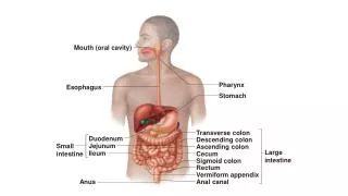
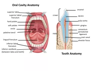
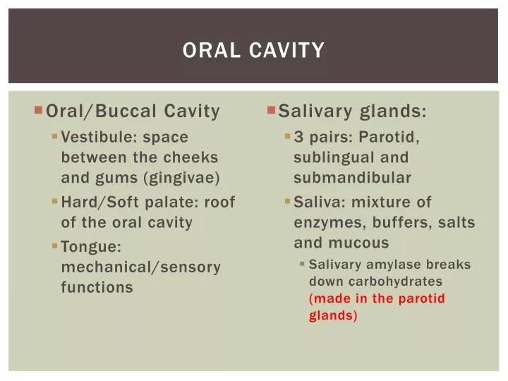
Oral cavity. Salivary glands: 3 pairs: Parotid , sublingual and submandibular Saliva: mixture of enzymes, buffers, salts and mucous Salivary amylase breaks down carbohydrates (made in the parotid glands). Oral/ Buccal Cavity Vestibule: space between the cheeks and gums (gingivae)

E N D
Oral cavity • Salivary glands: • 3 pairs: Parotid, sublingual and submandibular • Saliva: mixture of enzymes, buffers, salts and mucous • Salivary amylase breaks down carbohydrates (made in the parotid glands) • Oral/Buccal Cavity • Vestibule: space between the cheeks and gums (gingivae) • Hard/Soft palate: roof of the oral cavity • Tongue: mechanical/sensory functions
Pharynx AND ESOPHAGUS • Esophagus: Passageway between pharynx and stomach (~10in) • Swallowing (deglutition) of bolus (food) begins voluntarily at the mouth and continues involuntarily at the pharynx • Lower esophageal sphincter contracts to prevent backflow • Pharynx serves as a passageway for air and food • Divided into three parts: • Nasopharynx • Oropharynx • Laryngopharnyx
Swallowing (deglutition) • Voluntary Oral Phase: bolus moves against hard palate and tongue towards the pharynx • Involuntary Pharyngeal Phase: sensory receptors initiate the swallowing reflex; soft palate raises and epiglottis closes, directing bolus toward esophagus (food in trachea = choking) • Involuntary Esophageal Phase: bolus moves toward stomach by peristalsis *depending on type of food, process can take from 1-10sec
Clinical note: Esophagitis & Diaphragmatic (Hiatal) Hernias • Esophagitis: caused by a weak/relaxed lower esophageal sphincter (“heartburn”, GERD) • Hiatal Hernias: abdominal organs pushed into thoracic cavity through the esophageal hiatus.
The Anatomy of the stomach • Four regions: • Cardia • Fundus • Body • Pylorus • Pylorus sphincter • Histology • Musclarisexterna: circular, longitudinal, oblique • Mucosa: rugae are wrinkles in the mucosa; flattened when full *capacity of 1-1.5 liters *Greater and lesser omentum are mesenteries that protect and stabilize stomach(Fig. 16-8b)
Function and regulation of the stomach • Regulation • Cephalic Phase: sensory input accelerates production of gastric juice • Gastric Phase: food enters stomach and initiates contractions and secretions • Intestinal Phase: regulates flow of chyme into the duodenum • Functions: • Stores ingested food • Churns food and forms chyme • Secretes gastric juice (HCl, mucus, intrinsic factor, pepsinogen)
The small intestine • Anatomy • ~20ft long • 3 segments: • Duodenum: outside of peritoneum (10in) • Jejunum: inside peritoneum, bulk of absorption (8ft) • Ileum: longest segment, ileocecal valve controls flow of food into cecum (12ft)
The small intestine • Histology • Plicae (ply-see): folds along the intestinal lining • Villi: fingerlike projections contain microvilli (increase surface area and absorption into bloodstream) • Function • 90% absorption • Segmentation and breakdown of food by enzymes from accessory organs
homework • Chapter Objectives 1-5 (p. 515) • Vocabulary: mucosa; submucosa; muscularisexterna; serosa; peristalsis • Due: Wednesday, 4/10 • Chapter Objectives 6-11 • Due: Monday, 4/15 • Reading for tonight: pp. 523-536 (end at “the large intestine”)