Download
1 / 75
750 likes | 935 Views
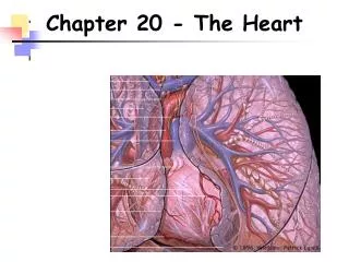
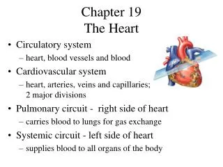
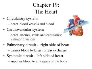
Chapter 19 The Heart. Circulatory system heart, blood vessels and blood Cardiovascular system heart, arteries, veins and capillaries; 2 major divisions Pulmonary circuit - right side of heart carries blood to lungs for gas exchange Systemic circuit - left side of heart

E N D
Chapter 19 The Heart • Circulatory system • heart, blood vessels and blood • Cardiovascular system • heart, arteries, veins and capillaries; 2 major divisions • Pulmonary circuit - right side of heart • carries blood to lungs for gas exchange • Systemic circuit - left side of heart • supplies blood to all organs of the body
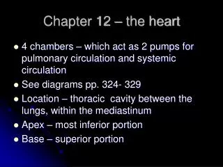
Size, Shape and Position • Located in mediastinum, between lungs • Base - broad superior portion of heart • Apex - inferior end, tilts to the left, tapers to point • 3.5 in. wide at base, 5 in. from base to apex and 2.5 in. anterior to posterior; weighs 10 oz
Pericardium • Allows heart to beat without friction, room to expand and resists excessive expansion • Parietal pericardium • outer, tough, fibrous layer of CT • inner, thin, smooth, moist serous layer • Pericardial cavity • filled with pericardial fluid • Visceral pericardium (a.k.a. epicardium of heart wall) • thin, smooth, moist serous layer covers heart surface
Heart Wall • Epicardium (a.k.a. visceral pericardium) • serous membrane covers heart • Myocardium • thick muscular layer • fibrous skeleton - network of collagenous and elastic fibers • provides structural support • attachment for cardiac muscle • nonconductor important in coordinating contractile activity • Endocardium • smooth inner lining
Pericardium & Heart Wall • Pericardial cavity contains 5-30 ml of pericardial fluid
Heart Chambers • 4 chambers • Right and left atria • 2 superior, posterior chambers • receive blood returning to heart • Right and left ventricles • 2 inferior chambers • pump blood into arteries • Atrioventricular sulcus - separates atria, ventricles • Anterior and posterior sulci - grooves separate ventricles (next slide)
External Anatomy - Anterior Atrioventricular sulcus
Heart Chambers - Internal • Interatrial septum • wall that separates atria • Pectinate muscles • internal ridges of myocardium in right atrium and both auricles • Interventricular septum • wall that separates ventricles • Trabeculae carneae • internal ridges in both ventricles
Heart Internal Anatomy • Heart bisected in frontal plane, opened like a book
Heart Valves • Ensure one-way blood flow • Atrioventricular (AV) valves • right AV valve has 3 cusps (tricuspid valve) • left AV valve has 2 cusps (mitral,bicuspid valve) lamb • chordae tendineae - cords connect AV valves to papillary muscles (on floor of ventricles) • Semilunar valves - control flow into great arteries • pulmonary: from right ventricle into pulmonary trunk • aortic: from left ventricle into aorta
AV Valve Mechanics • Ventricles relax, pressure drops, semilunar valves close, AV valves open, blood flows from atria to ventricles • Ventricles contract, AV valves close(papillary m. contract and pull on chordae tendineae to prevent prolapse),pressure rises, semilunar valves open, blood flows into great vessels
Coronary Circulation • Blood vessels nourish cardiac muscle • Left coronary artery • anterior interventricular artery • supplies interventricular septum + anterior walls of ventricles • circumflex artery • passes around left side of heart in coronary sulcus, supplies left atrium and posterior wall of left ventricle • Right coronary artery • marginal artery • supplies lateral R atrium + ventricle • posterior interventricular artery • supplies posterior walls of ventricles
Myocardial Infarction • Sudden death of heart tissue caused by interruption of blood flow from vessel narrowing or occlusion • Anastomoses defend against interruption by providing alternate blood pathways • circumflex artery and right coronary arterycombine to form posterior interventricular artery • anterior and posterior interventricular arteriescombine at apex of heart
Venous Drainage • 20% drains directly into right ventricle • 80% returns to right atrium • great cardiac vein (anterior interventricular sulcus) • middle cardiac vein (posterior sulcus) • coronary sinus (posterior coronary sulcus) collects blood from these and smaller veins and empties into right atrium
Coronary Flow and the Cardiac Cycle • Reduced during ventricular contraction • arteries compressed • Increased during ventricular relaxation • openings to coronary arteries, just above aortic semilunar valve, fill as blood surges back to valve
Structure of Cardiac Muscle • Short, thick, branched cells, 50 to 100 m long and 10 to 20 m wide with one central nucleus • Sarcoplasmic reticulum, large T tubules • must admit more Ca2+ from ECF during excitation • Intercalated discs, join myocytes end to end • interdigitating folds - surface area • mechanical junctions tightly join myocytes • fascia adherens: actin anchored to plasma membrane • desmosomes • electrical junctions - gap junctions form channels allowing ions to flow directly into next cell
Metabolism of Cardiac Muscle • Aerobic respiration • Rich in myoglobin and glycogen • Large mitochondria • Organic fuels: fatty acids, glucose, ketones • Fatigue resistant
Cardiac Conduction System • Myogenic - heartbeat originates within heart • Autorhythmic – regular, spontaneous depolarization • Conduction system • SA node: pacemaker, initiates heartbeat, sets heart rate • fibrous skeleton insulates atria from ventricles • AV node: electrical gateway to ventricles • AV bundle: pathway for signals from AV node • Right and left bundle branches: divisions of AV bundle that enter interventricular septum and descend to apex • Purkinje fibers: upward from apex spread throughout ventricular myocardium
Cardiac Rhythm • Systole = contraction; diastole = relaxation • Sinus rhythm • set by SA node, adult at rest is 70 to 80 bpm • Ectopic foci - region of spontaneous firing (not SA) • nodal rhythm - set by AV node, 40 to 50 bpm • intrinsic ventricular rhythm - 20 to 40 bpm • Arrhythmia - abnormal cardiac rhythm • heart block: failure of conduction system • bundle branch block • total heart block (damage to AV node)
Depolarization of SA Node • SA node - no stable resting membrane potential • Pacemaker potential • gradual depolarization from -60 mV, slow influx of Na+ • Action potential • occurs at threshold of -40 mV • depolarizing phase to 0 mV • fast Ca+2 channels open, (Ca+2 in) • repolarizing phase • K+ channels open, (K+ out) • at-60 mV K+ channels close, pacemaker potential starts over • Each depolarization creates one heartbeat • SA node at rest fires at 0.8 sec, about 75 bpm
Impulse Conduction to Myocardium • SA node signal travels at 1 m/sec through atria • AV node slows signal to 0.05 m/sec • thin myocytes with fewer gap junctions • delays signal 100 msec, allows ventricles to fill • AV bundle and purkinje fibers • speeds signal along at 4 m/sec to ventricles • Papillary muscles - get signal first, contraction stabilizes AV valves • Ventricular systole begins at apex, progresses up • spiral arrangement of myocytes twists ventricles slightly
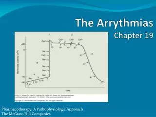
Contraction of Myocardium • Myocytes have stable resting potential of -90 mV • Depolarization(very brief) • stimulus opens voltage regulated Na+ gates, (Na+ rushes in) membrane depolarizes rapidly • action potential peaks at +30 mV • Na+ gates close quickly • Plateau - 200 to 250 msec, sustains contraction • slowCa+2 channels open, Ca+2 binds to fast Ca+2 channels on SR, releases Ca+2 into cytosol:contraction • Repolarization - Ca+2 channels close, K+ channels open, rapid K+ out returns to resting potential
Myocardial Contraction& Action Potential 1) Na+ gates open 2) Rapid depolarization 3) Na+ gates close 4) Ca+2 channels open 5) Ca+2 channels close K+ channels open
Electrocardiogram (ECG) • Composite of all action potentials of nodal and myocardial cells detected, amplified and recorded by electrodes on arms, legs and chest
ECG • P wave • SA node fires, atrial depolarization • atrial systole • QRS complex • AV node fires, ventricular depolarization • ventricular systole • (atrial repolarization and diastole - signal obscured) • T wave • ventricular repolarization
Electrical Activity of Myocardium 1) atrialdepolarization begins 2) atrial depolarization complete (atria contracted) 3) ventriclesbegin to depolarize at apex; atriarepolarize (atria relaxed) 4) ventricular depolarization complete (ventricles contracted) 5) ventriclesbegin torepolarize at apex 6) ventricular repolarization complete(ventricles relaxed)
Diagnostic Value of ECG • Invaluable for diagnosing abnormalities in conduction pathways, MI, heart enlargement and electrolyte and hormone imbalances
ECGs, Normal & Abnormal No P waves
ECGs, Abnormal Extrasystole : note the inverted QRS complex, misshapen QRS and T and absence of a P wave preceding this contraction.
ECGs, Abnormal Arrhythmia: conduction failure at AV node No pumping action occurs
Cardiac Cycle • One complete contraction and relaxation of all 4 chambers of the heart • Atrial systole, Ventricle diastole • Atrial diastole, Ventriclesystole • Quiescent period
Principles of Pressure and Flow • Measurement: force (mmHg) required to stop flow - sphygmomanometer • Change in volume creates a pressure gradient • Opposing pressures • great vessels have positive blood pressure • ventricular pressure must rise above this pressure for blood to flow into great vessels
Heart Sounds • Auscultation - listening to sounds made by body • First heart sound (S1), louder and longer “lubb”, occurs with closure of AV valves • Second heart sound (S2), softer and sharper “dupp” occurs with closure of semilunar valves • S3 - rarely heard in people > 30