Download
1 / 21
210 likes | 359 Views
Chapter 18: The Heart. Size, location, & orientation. Heart is about 250-350 grams (less than 1 lb) About the size of a persons fist Location – found in the mediastinum – medial cavity of thorax 2/3 of mass is left of midsternal Rests on superior surface of the diaphragm

E N D
Size, location, & orientation • Heart is about 250-350 grams (less than 1 lb) • About the size of a persons fist • Location – • found in the mediastinum – medial cavity of thorax • 2/3 of mass is left of midsternal • Rests on superior surface of the diaphragm • Anterior to the vertebral column • Posterior to the sternum • Flanked by the lungs • Base – • Posterior surface (top) • Broad & flat • 9 cm wide • Directed towards right shoulder • Apex – • Pointed end • Base of both ventricles • Point towards the left hip
Coverings • Heart is enclosed in the pericardium: • Protects the heart • Anchors it to surrounding structures (diaphragm) • Prevents over filling • Serous pericardium: • Thin serous membrane • Two layers: • Parietal – • Lines the inside of the pericardium – internal surface • Tough fibrous connective tissue layer • Anchors to diaphragm & sternum • Visceral – • Covers the external surface of the heart • Serous layer turns downward to cover the heart • Part of heart wall – laced with fat • Pericardial cavity – • Potential space – space between visceral & serous layer of parietal pericardium • Contains pericardial fluid – allows serous membranes to glide past one another
Layers of the heart wall • 3 layers: • Superficial epicardium (viseral pericardium): • Visceral layer of the serious pericardium • Infiltrated with fat • Middle myocardium: • Mostly cardiac muscle – bulk of the heart • Layer that actually contracts • Branching cardiac cells held together by crisscrossing connective tissue fibers & arranged in bundles • Connective tissue fibers form network – fibrous skeleton – reinforcement of heart muscle especially around valve areas & where vessels attach to the heart • Deep endocardium: • White sheet of endothelium resting on connective tissue layer • Located – inner myocardial surface, lining of chambers, & covers connective tissue skeleton of valves • Continuous w/ the endothelial linings of blood vessels leaving & entering the heart
Chambers • 4 chambers – 2 atria & 2 ventricles • Internal septum – • divides the atria internally longitudinally • Interventricular septum – • divides the ventricle internally longitudinally • Right ventricle = • most of the anterior surface • Left ventricle = • inferoposterior aspect & forms most of the apex
Chambers • Atria – • Superior • Receiving chambers for blood returning to the heart from circulation • Small chambers • Push blood to ventricles • Auricles – ear-like flap – from outside of each atria – increase atrial volume • Internal walls have ridges of muscles – pectinate • Fossa ovalis – shallow depression in interatrial septum – residual from fetal heart • Blood enters the right atrium from – • Superior vena cava – returns blood from above the diaphragm (upper body) • Inferior vena cava – blood returning from below the diaphragm (below the heart) • Coronary sinus – collects blood draining from myocardium • Blood enters the left atrium from – • 4 pulmonary veins • Seen from posterior view of the heart • Transport blood from the lungs
Chambers • Ventricles – • Inferior • Most of the mass of the heart • Right ventricle = anterior surface • Left ventricle = apex • Trabecuae carneae – • irregular ridges of muscles • Papillary muscles – • project into the ventricular cavity – play a role in valve function • (muscle folds = trabeculae carnae – some are stalklike and attach to valves = papillary muscles) • Discharging chambers – pumps of the heart • Blood propels out of the heart into circulation • Walls much thicker than atrial walls • Right ventricle – pulmonary trunk – routs blood to lungs (gass exchange occurs) • Left ventricle - aorta – body
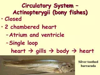
Blood pathway through heart • Pulmonary circuit – • Right side of the heart • Low pressure • Blood to and from the lungs • Blood flow path- • Blood in right atrium from body (oxygen poor, CO2 rich) via superior and inferior venae cavae • right ventricle • pumps into lungs (for oxygen pickup) via pulmonary trunk • from lungs to heart through pulmonary veins • left atrium • left ventricle
Blood pathway cont • Systemic circuit – • Left side of the heart • High pressure • Supplies body w/ oxygenated blood • Pathway – • From lungs • Pulmonary veins • Left atrium • Left ventricle – contracts • Aorta • Body
Valves • Atrioventricular valves (AV) – • Atrial-ventricular junction • Prevent backflow into the atria when ventricles are contracting • Attached to AV valves – collagen cords – chordae tendineane – anchor papillary muscles protruding from ventricular walls • Right AV valve = tricuspid valve • 3 flaps (tri) – endocardium & connective tissue • Left AV valve = bicuspid valve • 2 flaps (bi) • How it works – • When heart is relaxed – AV valves hang into ventricle • Blood into atria & into ventricle (through open AV valve) • Ventricle contracts • Ventricular pressure rises – forces blood (superiorly) against AV valve • Valve edges meet – closing the valve • Chordae tendineae & papillary muscles – anchor valves while they are closed
Valves • Semilunar (half moon) valves – • Major arteries leaving the heart • Prevent backflow into the ventricles • Aortic semilunar – • Valve at the base of the aorta • Pulmonary semilunar – • Valve at the base of the pulmonary trunk • No chordae tendinae – valve movement caused by force of blood • Heart is relaxed – valves are closed • Heart contracts – valves are forced open
Microscopic anatomy • Cardiac muscle – • Striated, short fat, branched, & interconnected muscle • One or two nuclei • Intracellular space filled w/ loose connective tissue matrix & capillaries – connected to fibrous skeleton – allows cardiac cells to exert force • Contraction occurs via sliding filaments • Adjacent muscles interconnect @ intercalated discs • Disc contains desmosomes (hold cardiac cells together) & gap junctions (allow ions to pass from cell to cell) • Allow cardiac cells to electrically behave as a unit • High concentration of mitochondria • Metabolize fatty acids for ATP • Can switch nutrient pathways to use whatever nutrient supply that is available • Depends on a continual supply of oxygen • Aerobic respiration – can’t have oxygen deprivation & still operate
Heart contraction • Sodium & calcium needs: • Sodium ions enter cardiac muscle cells from extracellular fluid (sodium ion channels) • Causes a depolarization that causes the sarcoplasmic reticulum (specialized ER) to release calcium • Calcium enters the sarcoplasm (cytoplasm of cardiac cells) • Calcium signals myofilaments (individual muscle fibers) to contract • Cardiac muscles contract as a unit or not at all
Electrical events • Intrinsic cardiac conduction system – made up of specialized cardiac cells –nodal system • Initiate & distribute impulses • Ensures that the heart depolarizes in sequential order • Contracts because of gap junction (allows signals to pass between cells)
Electrical events • Sequence of excitation – • 1. SA node – • Sinoatrial • Pacemaker - sets pace for the heart • In right atrial wall • Minute cell mass • Depolarizes 70-80 times per min • Called sinus rhythm – determines heart rate • 2. AV node – • Atrioventricular • In interatrial septum – above the tricuspid valve • Depolarization spreads via gap junctions • From SA node to AV node • Impulse delayed 1 sec to allow atria to completely contract • 3. AV bundle – • Atrioventricular • “Bundle of His” • Superior part of interventricular septum • Connects atria & ventricles electronically
Sequence of excitation cont. • 4. right & left bundles – • Along interventricular septum toward apex of the heart • Conduct signal through ventricles • 5. ventricular walls – • Penetrate the heart apex • Turn superiorly into ventricular walls • Bundle branches excite septal cells • Contraction depends on cell-to-cell transmission via gap junctions • Total elapsed time from SA node to ventricular node = .22 sec • Ventricles contract w/ wringing motion beginning at apex, moving toward atria
Excitation http://www.youtube.com/watch?v=MGxxRyJTmwU&feature=related
Heart sounds & CO • Lub – • 1st heart sound • AV valves close • Beginning of systole (ventricular pressure rises above atrial pressure) • Dup – • 2nd heart sound • Closure of semilunar valves • During ventricular diastole • Cardiac output (CO) – • Amount of blood pumped out each ventricle in one minute (one cycle) • CO = HR x SV • Stroke volume (SV) – • Volume of blood pumped by a ventricle with each beat (during one contraction) • Correlated w/ force of ventricular contraction
Defects • Murmurs – • Abnormal heart sounds • Indicate valve problems • Tachycardia – • Abnormally fast heart rate • Over 100 beats per min • Arrhythmias – • Uncoordinated contractions • Irregular heart rhythms • Fibrillation – • Rapid, irregular, out of phase contractions • Heart is useless • Must be defibrillated immediately before brain death (electric shock) • Abnormal pacemaker – • Ectopic pacemaker • AV node may take over – but at a slower pace • Caffeine can cause irregular rhythms separate from the SA node
Cardiac cycle • Ventricular filling – • Flows passively through atria into ventricles (70% of blood) • Atria contract propelling left over blood into ventricles • Ventricular systole – • Atria relax • Ventricles contract • Pressure rises closing the AV valves • Semilunar valves forced open • Blood passes into the aorta & pulmonary trunk • Isovolumetric relaxation – • Ventricular pressure drops • Closes the semilunar valves • Blood rebounds off the valves & continues its path