Download
1 / 17
170 likes | 294 Views
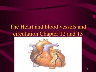
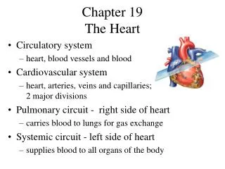
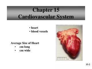
Chapter 12 – the heart. 4 chambers – which act as 2 pumps for pulmonary circulation and systemic circulation See diagrams pp. 324- 329 Location – thoracic cavity between the lungs, within the mediastinum Apex – most inferior portion Base – superior portion . Heart anatomy. diagram.

E N D
Chapter 12 – the heart • 4 chambers – which act as 2 pumps for pulmonary circulation and systemic circulation • See diagrams pp. 324- 329 • Location – thoracic cavity between the lungs, within the mediastinum • Apex – most inferior portion • Base – superior portion
Heart anatomy • diagram
Go here for the best diagrams • all kinds of wonderful information on the heart!
External anatomy • Coronary sulcus • Superior and inferior vena cava • Pulmonary veins • Pulmonary trunk • Aorta • Membranes: pericardial cavity, pericardium: fibrous, serous, parietal, visceral, pericardial fluid
Blood supply to the heart • Coronary arteries (left and right) • Cardiac veins • Coronary sinus
Heart chambers • Atria – right and left • Auricle • Interatrial septum • Ventricles – right and left • Interventricular septum
Heart valves • Atrioventricular valves • Tricuspid valve – between right atrium and right ventricle • Bicuspid (mitral) valve – between left atrium and laft ventricle • Papillary muscles • Chordae tendineae • Aortic and pulmonary semilunar valves
Blood flow through the heart • Both atria contract at the same time and both ventricles contract at the same time • Body vena cava right atrium tricuspid valve right ventricle pulmonary semilunar valve pulmonary arteries lungs pulmonary veins left atrium bicuspid valve left ventricle aortic semilunar valve aorta body
Heart wall • Three layers: • Epicardium • Myocardium • endocardium
Cardiac muscle • Striated • Elongated, branching cells • 1 or 2 nuclei • Rich in mitochondria • ATP • Intercalated disks
Conduction system of the heart • SA node • AV node • AV bundle • Left and right bundle branches • Purkinje fibers • See diagram pg. 336 • Ectopic beat
electrocardiogram • P wave • QRS complex • T wave • P-Q interval • P-R interval • Q-T interval
Cardiac cycle • Systole – atrial and ventricular – contraction • Diastole – atrial and ventricular – relaxation • Heart sounds – stethoscope • Murmurs • Incompetent valve • Stenosed opening
Regulation of heart function • Cardiac output • Stroke volume • Heart rate • Cardiac output = stroke volume x heart rate
Intrinsic regulation • Regulation mechanisms contained within the heart • Venous return • Preload • Starling’s law of the heart • afterload
Extrinsic regulation • Regulation by factors outside the heart – nervous and hormonal regulation • Baroreceptor reflex • Medulla oblongata – cardioregulatory center