Download
1 / 34
350 likes | 491 Views
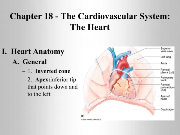
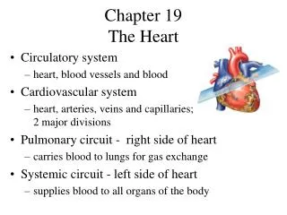
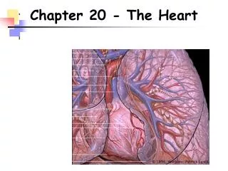
The Heart Chapter 18. The Heart . Size – 250-350 grams Location – in mediastinum of thoracic cavity Function – generates pressure to pump blood through circulatory system Orientation – flat base is directed toward right shoulder, and pointed apex points to left hip. Figure 18.1.

E N D
The Heart Chapter 18
The Heart • Size – 250-350 grams • Location – in mediastinum of thoracic cavity • Function – generates pressure to pump blood through circulatory system • Orientation – flat base is directed toward right shoulder, and pointed apex points to left hip Figure 18.1
Heart Coverings Pericardium – the two layered, membranous sac in which the heart sits Fibrous pericardium – the outer, thick layer composed of dense connective tissue, for protecting, anchoring the heart in position, and preventing overfilling Serous pericardium – the inner thin layer composed of a serous membrane Visceral layer – membrane clinging to the outer heart surface Parietal layer – membrane clinging to the inside of the fibrous pericardium Pericardial cavity – serous fluid-filled space between the visceral and parietal layers Figure 18.2
Heart Layers Epicardium – the epithelium clinging to the outer heart wall (is the visceral pericardium) Myocardium – the middle layer composed of cardiac muscle tissue Endocardium – the epithelium clinging to the inner surfaces of the heart chambers Figure 18.2
The Heart • 4 chambers: 2 atria, 2 ventricles • Atria : • the superior chambers • auricles – ear-like extensions of the atria • receiving chambers, limited pumping means thin walls • Ventricles: • the inferior chambers, the majority of the heart volume • pumping chambers, thick walls • Sulci: • The indentations on the outer heart surface, corresponding to borderlines between chambers • contain fat and vessels • Ex – interventricular sulcus Figure 18.4 a, b
The Heart • Septa: • The internal walls that divide the chambers • Correspond to the external sulci • Ex’s- Interventricular septum and Interatrial septum Figure 18.4e, f
The Atria • Right Atrium Entrances: • Superior Vena Cava – blood returning from above the diaphragm • Inferior Vena Cava – blood returning from below the diaphragm • Coronary sinus – blood returning from the heart wall • Left Atrium Entrances: • 4 pulmonary veins - blood returning from lungs Figure 18.4e
The Ventricles • Right Ventricle: • Receives blood from the right atrium • Blood exits into the pulmonary trunk – to lungs • Left Ventricle: • Receives blood from the left atrium • Blood exits into the aorta – to the body Figure 18.4e
Circulation • Blood only passes through ½ of the heart at a time, and therefore must pass through the heart twice to complete circulation • Pulmonary circuit: • the pathway from the heart to the lungs and back • is pumped by the right half of the heart • blood leaves –O2 and returns +O2 • Systemic circuit: • the pathway from the heart to the body’s tissues and back • is pumped by the left half of the heart • blood leaves +O2 and returns –O2 • NOTE – arteries do NOT always carry oxygenated blood, and veins do NOT always carry deoxygenated blood Figure 18.5
Circulation • The Systemic circuit is a longer circuit than the Pulmonary circuit • Greater pressure is needed to pump blood through the systemic circuit • The left ventricle therefore has more cardiac muscle tissue than the right ventricle Figure 18.6
Coronary Circulation Coronary circulation – the series of vessels that supply blood flow to the wall of the heart, beginning at the aorta and ending at the right atrium Anastomoses – the merging of blood vessels, providing more than one way to deliver blood to one location…Why? Myocardial Infarction – “heart attack” – due to a blockage of a coronary artery, causing the death of cardiac muscle cells Figure 18.7
Coronary Circulation Coronary arteries – deliver oxygen and nutrient rich blood to the cardiac muscle Figure 18.4b
Coronary Circulation Cardiac veins – drain the oxygen and nutrient poor blood from the cardiac muscle Coronary sinus – large vein on posterior side that empties into the right atrium Figure 18.4d
Heart Valves Function – ensure a unidirectional flow of blood through the heart Types – there are 2 atrioventricular valves and 2 semilunar valves Figure 18.8 a, b
Heart Valves • Atrioventricular valves: • separate an atrium from a ventricle • prevent backflow into the atrium • Tricuspid (Right AV) valve – separates right atrium from right ventricle • Bicuspid (Left AV) valve – separates left atrium from left ventricle. Also known as mitral valve • Flaps (cusps) of these valves are supported by papillary muscles and chordae tendineae Figure 18.8c
Heart Valves • Atrioventricular valves: • Papillary muscles attach to valve flaps via chordae tendineae • These muscles contract to prevent the valve flaps from inverting into the atria Figure 18.9
Heart Valves • Semilunar valves: • separate a ventricle from a great vessel • prevent backflow into the ventricle • composed of three cup-like flaps • Pulmonary semilunar valve – regulates movement of blood into pulmonary trunk • Aortic semilunar valve – regulates movement of blood into the aorta • Flaps (cusps) of these valves are NOT supported by papillary muscles and chordae tendineae Figure 18.8c
Heart Valves • Semilunar valves: • during contraction, pressure forces these valves open, allowing blood into the large vessels • when the ventricles relax, the blood falls toward the ventricles, fills the cup-shaped flaps, closing the valves Figure 18.10
Microscopic Anatomy • Cardiac muscle tissue: • Striated – a striped appearance • Involuntary – no conscious control • Cardiac Muscle Fibers (cells): • shorter than skeletal muscle fibers, with central nuclei • large amount of mitochondria for endurance • branched – fibers divide and unite • interconnected – fibers are linked and work in unison Figure 18.11
Cardiac Muscle • Cardiac Muscle Fibers (cells): • Intercalated discs- junctions where adjacent fibers connect • desmosomes – hold fibers tightly together • gap junctions – allow fibers to share cytoplasm and contract in unison Figure 18.11a
Cardiac Muscle Contraction Autorhythmicity The ability of cardiac muscle to trigger its own contraction, not needing a nervous system impulse Whole organ contraction There is no partial contraction of cardiac muscle, it is an all or none event Figure 18.11
Cardiac Muscle Contraction • Action potentials in cardiac muscle involve 3 ions (Na+, K+ and Ca2+), which produces a plateau phase of the impulse • This prolongs the contraction to ensure efficient blood ejection • Also increases the time of the absolute refractory period, ensuring separate heart contractions (not prolonged muscle tetanus) Figure 18.12
Intrinsic Conduction System • Intrinsic Conduction System: • Stimuli that trigger cardiac muscle contraction come from within the heart itself • Autorhythmic cells - specialized heart cells that generate action potentials which spread through the heart to trigger contraction • These cells have unstable resting potentials, and therefore depolarize regularly • These pacemaker potentials cause action potentials in the cardiac muscle fibers, triggering contraction Figure 18.13
Intrinsic Conduction System Sinoatrial node – in the right atrium, the “pacemaker” whose cells generate the sinus rhythm of 75 beats/min Atrioventricular node - causes a delay of .1sec to allow atrial contraction before ventricular contraction. Rhythm is 40-60 beats/min Bundle of His – pathway into the interventricular septum Figure 18.14
Intrinsic Conduction System • Bundle Branches – the right and left pathways through the interventricular septum • Purkinje fibers – pathways to the walls of the ventricles • The bundle of his, bundle branches and purkinje fibers would set a rhythm of only 30 beats/min Figure 18.14
Extrinsic Innervation Cardiac centers – gray matter areas in the medulla that can change heart rhythm Parasympathetic innervation – via the vagus nerve, slows heart rate Sympathetic innervation – via the sympathetic trunk, increases heart rate Heart rate disorders: Tachycardia– an abnormally fast heart rate 100+ beats/min Bradycardia – an abnormally low heart rate 60- beats /min Arrhythmias – irregular heart rhythms Fibrillation – rapid, irregular contractions that do not function to pump blood Figure 18.15
Electrocardiogram ECG ECG – a graphic representation of all of the action potentials in the heart in a given time P wave – shows the depolarization of the atria QRS complex – shows the depolarization of the ventricles T wave – shows the repolarization of the ventricles Figure 18.16
Electrocardiogram ECG Note: the waves of the ECG graph correspond to the spreading depolarization through the heart tissue. Muscle contraction follows this depolarization Figure 18.17
Electrocardiogram ECG Figure 18.18
Heart Sounds Heart sounds – the “lub dub” “Lub” – the sound produced by the closure of the AV valves “Dub” – the sound produced by the closure of the semilunar valves Murmurs – abnormal heart sounds, indicating valve problems Figure 18.19
Cardiac Cycle • Cardiac cycle – the events of a single heart beat • Systole – heart contraction Diastole – heart relaxation • Ventricular filling – blood flows through the atria into the ventricles. The AV valves are open, and the semilunar valves are closed. Then, the atria contract forcing the remaining blood into the ventricles • Isovolumetric contraction – ventricles contract, forcing the AV valves closed. The volume in the ventricles is now the end diastolic volume (EDV) Figure 18.20
Cardiac Cycle 3. Ventricular ejection – ventricular pressure forces the semilunar valves open and the blood enters the great vessels 4. Isovolumetric relaxation – ventricles relax, and the blood within them is the end systolic volume (ESV). The semilunar valves close and the atria begin to fill. 5. Ventricular filling – atrial pressure forces the AV valves open which restarts the cycle Figure 18.20
Cardiac Output Cardiac Output (CO) – the amount of blood pumped by each ventricle in one minute CO = heart rate x stroke volume Stroke volume (SV) = EDV - ESV Practice Question: If a person’s EDV is 125mL and their ESV is 50mL, what is their CO if their heart rate is 80 beats/min? Answer: SV = 125mL – 50 mL SV = 75mL CO = 75mL x 80 beats/min CO = 6000mL/min = 6.0L/min Figure 18.21
Fetal Heart Structures Foramen ovale – a hole in the interatrial septum allowing blood to pass from the right atrium to the left atrium. Its remnant is the fossa ovalis in adults Ductus arteriosus – a passage from the pulmonary trunk to the aorta. Its remnant is the ligamentum arteriosum in adults Both structures allow the fetal blood to bypass the pulmonary circuit….Why? Figure 18.24