Download
1 / 24
270 likes | 438 Views
Scanning electron microscopy. Tracy Furutani November 13, 2008. Uses photons of visible light Resolution 1000 nm Color images Lenses of glass Aberration in lenses difficult to correct Sample in air. Uses electrons Resolution 1 nm Color meaningless Lenses of solenoid coils

E N D
Scanning electron microscopy Tracy Furutani November 13, 2008
Uses photons of visible light Resolution 1000 nm Color images Lenses of glass Aberration in lenses difficult to correct Sample in air Uses electrons Resolution 1 nm Color meaningless Lenses of solenoid coils Stigmator allows control of beam Sample in vacuum Light microscope vs SEM
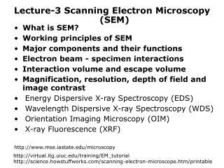
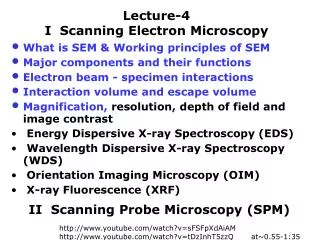
Types of electron microscopy • Transmission electron microscopy -- first invented (1931), works like light microscope but with electrons, needs thin samples • Scanning electron microscopy -- have beam of electrons “scan” across a surface to image it • Reflection electron microscopy -- detects the pattern of scattered electrons • Scanning transmission electron microscopy -- combining SEM sample interaction with TEM imaging • Scanning tunneling microscopy -- electrons “tunnel” through vacuum to “feel” surface
Some definitions • Stigmation -- correcting asymmetries in horizontal versus vertical focus. If the image has astigmatism there will be “streakiness” in the image. • Collimation -- the creation of parallel path particles (in this case, electrons)
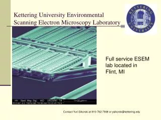
Sample preparation • Coated (sputtered) versus noncoated • Stubs versus vises • Carbon tape versus carbon paint
Some techniques to improve performance • Spot size -- literally the size of the path of the focused electrons as they strike the sample • Minimize this to improve focus at high magnification by a) decreasing the working distance or b) increasing the current on the focusing lens • Trade off: smaller area of coverage, lower beam current means worse contrast
More techniques to improve performance • Depth of field -- how much relief can be on the sample with it still staying in focus • Maximizing this allows you to see detail on rough surfaces by a) decreasing the aperture size, b) decreasing the magnification or c) increasing the working distance • Trade off: lower magnification
Even more techniques to improve performance • Signal-to-noise ratio -- the contrast between interacting and noninteracting surfaces • Maximize this to gain more fine detail by a) using a high beam current or b) using a slower scan rate • Trade off: Much larger spot size
Energy dispersive X-ray spectroscopy • Known as EDS, EDX or EDXRF • Used for elemental analysis • Electrons from SEM beam excite electrons in atoms in sample • Atoms in sample emit characteristic energy X-rays as electrons drop back to ground state