Download
1 / 34
410 likes | 629 Views
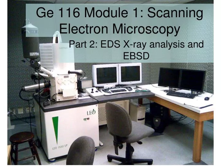
Ge 116 Module 1: Scanning Electron Microscopy. Part 2: EDS X-ray analysis and EBSD. Continuum X-rays. Characteristic X-rays. Characteristic X-rays. Characteristic X-rays. X-ray counting: EDS and WDS. X-ray counting: EDS and WDS.

E N D
Ge 116 Module 1: Scanning Electron Microscopy Part 2: EDS X-ray analysis and EBSD
X-ray counting: EDS and WDS • Spectral resolution determined by electron-hole pair production energy and thermal noise
X-ray counting: EDS and WDS • Silicon Drift Detector (SDD) – new! • Low capacitance allows MUCH higher counting rate • Reaches optimal resolution at higher temperature (LN2 not required!)
X-ray counting: EDS and WDS • Rise time of steps depends on capacitance of system, limits counting rate. • Conventional Si detector is periodically discharged. SDD is continuously discharged (less dead time).
Complexities in X-ray production • Production, (z)
Complexities in X-ray production • Absorption
Complexities in X-ray production • Absorption
Complexities in X-ray production • Secondary Fluorescence
Complexities in X-ray production • Secondary Fluorescence 100 m From Milman-Barris et al. (2008)
Complexities in X-ray production • Quantitative analysis requires correction for production, absorption, and fluorescence effects • Physics-based methods: ZAF, (z) • Empirical method: Bence-Albee • Correction depends on composition, which is not known a priori, so quantification is an iterative procedure • Accurate analysis requires appropriate standards, as we will see when we learn electron probe analysis
Diffraction: Bragg Equation • where n is an integer, λ is the wavelength of the electrons, d is the spacing of the diffracting planes, and θ is the angle of incidence of the electrons on the diffracting plane • Constructive interference between reflections off successive planes of charge in the lattice requires difference in path length to be an integer multiple of the wavelength.
Aside: X-ray Diffraction • X-ray diffraction is usually done with a plane-wave X-ray source • For monochromatic X-radiation and a single crystal, this gives a distribution of points of constructive interference around the sphere. • For monochromatic X-radiation and a powdered material, this gives a set of single cones with opening angle 2 around the irradiation vector. • For white incident X-ray source and powdered material, energy-dispersive detector at fixed 2 angle sees a set of discrete energy peaks
Aside: X-ray Diffraction • X-ray diffraction is usually done with a plane-wave X-ray source • For monochromatic X-radiation and a single crystal, this gives a distribution of points of constructive interference around the sphere. • For monochromatic X-radiation and a powdered material, this gives a set of single cones with opening angle 2 around the irradiation vector. • For white incident X-ray source and powdered material, energy-dispersive detector at fixed 2 angle sees a set of discrete energy peaks
Aside: X-ray Diffraction • X-ray diffraction is usually done with a plane-wave X-ray source • For monochromatic X-radiation and a single crystal, this gives a distribution of points of constructive interference around the sphere. • For monochromatic X-radiation and a powdered material, this gives a set of single cones with opening angle 2 around the irradiation vector. • For white incident X-ray source and powdered material, energy-dispersive detector at fixed 2 angle sees a set of discrete energy peaks So, for 10 keV, 1.24 angstroms
Kikuchi pattern formation • (Observed in TEM in 1928!) So, for 10 keV, 0.124 angstroms
Kikuchi pattern formation • The monument to Kikuchi in Kumamoto (?)
Band detection • Hough Transform • 5 to 7 lines is usually enough for phase ID and orientation
Pattern indexing • Good pattern match determines crystal structure and orientation
EBSD experiment modes • Point analysis: phase and orientation determined at each analytical point
EBSD experiment modes • Orientation mapping
EBSD experiment modes • Grain mapping
EBSD experiment modes • Texture
EBSD experiment modes • Phase discrimination (automated point counting!)