Download
1 / 8
80 likes | 242 Views
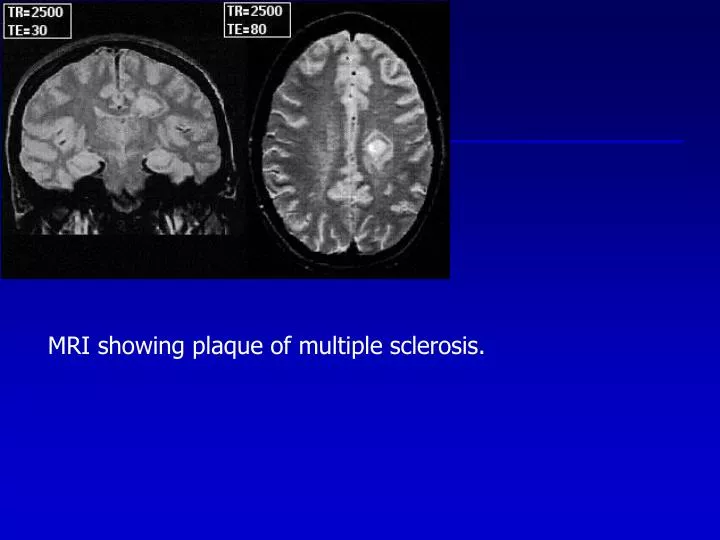
MRI showing plaque of multiple sclerosis. Left: Coronal section of cerebrum showing multiple sclerosis Plaques at the apex of both lateral ventricles. Right: HE/LFB stain of axial section of pons showing several Multiple sclerosis plaques. Left : Sharp edge of demyelination in MS plaque.

E N D
Left: Coronal section of cerebrum showing multiple sclerosis Plaques at the apex of both lateral ventricles. Right: HE/LFB stain of axial section of pons showing several Multiple sclerosis plaques.
Left: Sharp edge of demyelination in MS plaque. Right: Macrophage infiltrate and astrogliosis in MS plaque.
Axial section of pons and cerebellum showing discoloration And softening of central pontine myelinolysis.
Axial whole-mount section of cerebrum stained with LFB Showing caudal-to-rostral gradient of demyelination in adrenoleukodystrophy.
Loss of myelin and perivascular inflammatory infiltrate in affected Region of white matter in adrenoleukodystrophy (HE stain).
Cerebral white matter showing Globoid cells of Krabbe’s disease (HE stain).
Subpial collection of Rosenthal fibers in a cerebral gyrus from an infant who died of Alexander’s disease.