Download

1 / 14
140 likes | 292 Views
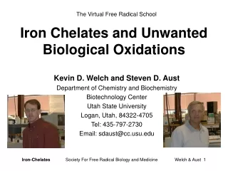
The Virtual Free Radical School. Iron Chelates and Unwanted Biological Oxidations. Kevin D. Welch and Steven D. Aust Department of Chemistry and Biochemistry Biotechnology Center Utah State University Logan, Utah, 84322-4705 Tel: 435-797-2730 Email: sdaust@cc.usu.edu. Introduction.

E N D
The Virtual Free Radical School Iron Chelates and Unwanted Biological Oxidations Kevin D. Welch and Steven D. Aust Department of Chemistry and Biochemistry Biotechnology Center Utah State University Logan, Utah, 84322-4705 Tel: 435-797-2730 Email: sdaust@cc.usu.edu Iron-ChelatesSociety For Free Radical Biology and Medicine Welch & Aust 1
Introduction Reed, C.A. (1985) in:The Biological Chemistry of Iron. (Dunford, H.B., Dolphin, D., Raymond, K.M., and Sieker, L., Eds.) D. Reidel, Boston, MA. • Versatility of oxidation state, reduction potential, coordination number, spin state, ligand type, ligand equilibria, ligand dynamics and structure give iron its special characteristics. • Iron is an integral biological cofactor for many proteins. Iron is used in many different types of cofactors, e.g., heme moieties, iron-sulfur clusters, di-iron centers, etc. • Iron presents a significant paradox to the field of oxygen radicals in biology. It is required for many cellular functions yet it can also catalyze the deleterious oxidation of biomolecules. Aust, S.D. and Koppenol, W.H. (1991) in: Oxidative Damage and Repair: Chemical, Biological and Medical Aspects (Davies, K.J.A., Ed.), pp. 802-807, Pergamon Press. Iron-ChelatesSociety For Free Radical Biology and Medicine Welch & Aust 2
Chelation of Iron • The reactivity of iron is highly dependent upon its ligand environment. For example, chelators that contain oxygen ligands tend to stabilize Fe(III), while chelators that contain nitrogen or sulfur ligands tend to stabilize Fe(II). • Ligation of iron by chelators that stabilize the ferrous form of iron, such as phenanthrolines, results in an increase in the reduction potential of the iron (+1.1 V). Conversely, ligation of iron by chelators that stabilize the ferric form of iron, such as deferroxamine, results in a decrease in the reduction potential of the iron ( -0.4 V). Miller, D.M., Buettner, G.R., and Aust, S.D. (1990) Free Radic. Biol. Med.8, 95-108. Iron-ChelatesSociety For Free Radical Biology and Medicine Welch & Aust 3
The Effect of pH and Buffers on the Rate of Fe(II) Autoxidation In general the rate of Fe(II) autoxidation in an aqueous solution (in the absence of any other chelators) is proportional to the square of [OH-]. rate = [Fe(II)][O2][OH-]2 Harris, D.C. and Aisen, P. (1973) Biochim. Biophys. Acta329, 156-158. However, relatively strong chelators such as phosphate, can somewhat override the effect of pH on the rate of Fe(II) autoxidation. For example, almost no Fe(II) autoxidizes in a HEPES buffered solution at pH 6.5, conversely, Fe(II) autoxidizes very rapidly in phosphate buffer, at pH 6.5. Welch, K.D., Davis, T.Z., and Aust, S.D. (2002) Arch. Biochem. Biophys.397, 360-369. Iron-ChelatesSociety For Free Radical Biology and Medicine Welch & Aust 4
The Effect of Chelation on the Rate and Stoichiometry of Fe(II) Autoxidation It has been shown experimentally that chelators, in general, affect the stability of Fe(II) as expected, i.e., chelators that ligate Fe(II) via oxygen ligands promote the autoxidation of Fe(II), whereas the autoxidation of Fe(II), ligated by chelators with nitrogen ligands, is slower. Interestingly, the stoichiometry of the autoxidation reaction appears to be inversely related to the rate, i.e., the faster the rate of Fe(II) autoxidation the lower the stoichiometry. Welch, K.D., Davis, T.Z., and Aust, S.D. (2002) Arch. Biochem. Biophys.397, 360-369. Iron-ChelatesSociety For Free Radical Biology and Medicine Welch & Aust 5
Mechanisms of Iron-Mediated Toxicity • Iron can mediate the deleterious oxidation of biomolecules, which can have serious consequences for an organism, resulting in various diseases, e.g., cancer, atherosclerosis, and diabetes. • Iron can oxidize numerous biomolecules indirectly via partially reduced oxygen species that can be produced in the presence of iron. One of the most commonly accepted mechanisms of iron-mediated oxygen radical production is described by the Haber-Weiss series of reactions shown below. • Iron can also oxidize numerous biomolecules via direct transfer of an electron from the molecule to an iron complex, e.g., ascorbate and dopamine. • [1] Fe (II) + O2 Fe (III) + O2- • [2] 2 O2 -+ 2H+ H2O2 + O2 • [3] H2O2 + Fe (II) Fe(III) + HO + OH- Reilly, C.A. and Aust, S.D. (2000) in: Toxicology of the Human Environment. The critical role of Free Radicals. C.J. Rhodes, Ed., Taylor & Francis Publ., London. pp 155-190. Iron-ChelatesSociety For Free Radical Biology and Medicine Welch & Aust 6
Fenton Reagent 50 mM PO4 Buffer 50 mM Tris Buffer PO4 + 100 U catalase Fe(II):EDTA 50 mM PO4 Buffer 50 mM Tris Buffer PO4 + 100 U catalase Tris + 100 U catalase Tris + 200 U catalase Welch, K.D., Davis, T.Z., and Aust, S.D. (2002) Arch. Biochem. Biophys.397, 360-369. Comparison of EtOH Oxidation by Fenton Reagent vs Fe:EDTA The autoxidation of some iron chelates results in the production of a strong oxidant, which is not the hydroxyl radical. The DMPO adduct trapped during the oxidation of EtOH by HO is different than the adduct trapped during the oxidation of EtOH by Fe(II):EDTA. Additionally, the oxidation of EtOH by the HO is inhibited by both the presence of organic buffers such as Tris and the addition of catalase. Whereas the oxidation of EtOH by Fe(II):EDTA is not inhibited by the presence of organic based buffers. The oxidation of EtOH by Fe(II):EDTA is sensitive to, but not dependent upon, H2O2. These data suggest that a ferryl iron species may be formed during the oxidation of Fe(II):EDTA. Iron-ChelatesSociety For Free Radical Biology and Medicine Welch & Aust 7
Direct Oxidation of Biomolecules Many biomolecules are good Fe(III) reductants, however, they are also good iron chelators. Therefore, they will reduce Fe(III) to Fe(II) and subsequently chelate the Fe(II), which can promote the oxidation of the Fe(II) to Fe(III) with the concomitant reduction of O2 to O2-. Thus, it appears as if the biomolecule autoxidized when in fact the oxidation of the biomolecule was mediated by a transition metal such as iron. The oxidation of ascorbate by iron is an excellent example. Buettner, G.R. (1988) J. Biochem. Biophys. Meth.16, 27-40. The oxidation of epinephrine has been shown to be catalyzed by contaminating metals. Any treatment of the solutions to remove or chelate transition metals resulted in an inhibition of the oxidation of epinephrine. Ryan, T.P., Miller, D.M., and Aust, S.D. (1993) J. Biochem. Toxicol.8, 33-39. Iron-ChelatesSociety For Free Radical Biology and Medicine Welch & Aust 8
Schematic Representation of Lipid Peroxidation The process of lipid peroxidation occurs via a free radical mechanism promoted by iron. The first step is the abstraction of a hydrogen atom, which results in the formation of a lipid radical. There are different theories as to how iron is involved in the initiation of lipid peroxidation. One hypothesis is that a Fe(II)-O2-Fe(III) species is required for iron-mediated lipid peroxidation. Bucher, J.R., Tien, M. and Aust, S.D. (1983) Biochem. Biophys. Res. Commun.111, 777-784. Iron can also breakdown existing lipid hydroperoxides, in a Fenton-like reaction, which will assist in the propagation steps of lipid peroxidation. Iron-ChelatesSociety For Free Radical Biology and Medicine Welch & Aust 9
The Dependence of Lipid Peroxidation on the Fe(II):Fe(III) Ratio It has been shown that maximal rates of lipid peroxidation are observed when the the ratio of Fe(II):Fe(III) is 1:1. Minotti, G. and Aust S.D. (1987) J. Biol. Chem. 262, 1098-1104. The addition of Fe(II), chelated by any one of a number of different iron chelators, in a liposomal system often results in a short lag period before lipid peroxidation starts. The addition of a second chelator results in an increase in the lag period or inhibition of lipid peroxidation. The degree of inhibition depends on the stability constant of the iron:chelator complex. The most pronounced effects were observed for the chelators with higher stability constants, which supports the hypothesis that a 1:1 ratio of Fe(II):Fe(III) is responsible for iron-mediated lipid peroxidation. Tang, L.X. et al. (1997) J. Inorg. Biochem.68, 265-272. Iron-ChelatesSociety For Free Radical Biology and Medicine Welch & Aust 10
Ascorbate (uM) The Effect of Ascorbic Acid Concentration on ADP:Fe(III)-dependent Lipid Peroxidation Ascorbic acid is able to promote the oxidation of lipids. The role of ascorbic acid in lipid peroxidation can be explained by the fact that ascrobate can reduce Fe(III) to give the 1:1 ratio of Fe(II):Fe(III). A high ratio of ascorbate to Fe(III) results in essentially all Fe(II), and less lipid peroxidation. Miller, D.M. and Aust, S.D. (1988) Arch. Biochem. Biophys.271, 113-119. Iron-ChelatesSociety For Free Radical Biology and Medicine Welch & Aust 11
Protein Oxidation • Molecular oxygen, Fe(III), and an appropriate electron donor can catalyze the oxidative modification of proteins. Only a few amino acids are modified and relatively little peptide bond cleavage occurs when proteins are exposed to iron-mediated oxidation systems. The available data indicate that iron-mediated oxidation systems catalyze the reduction of Fe(III) to Fe(II) and of O2 to H2O2 and that these products react at metal-binding sites on the protein to produce active strong oxidants (OH, ferryl ion) which attack the side chains of amino acid residues at the metal-binding site. • Iron-binding sites on proteins serve as centers for repeated production of OH that are generated via the Fenton reaction. Prevention of the site-specific free radical damage can be accomplished by using selective chelators for iron, by introducing high concentrations of HO scavengers, and by adding enzymes that remove O2- to H2O2. Histidine, for example, is a compound that can intervene in free radical reactions in a variety of modes. Stadtman, E.R. (1990) Free Radic. Biol. Med.9, 315-325. Chevion, M. (1988) Free Radic. Biol. Med.5, 27-37. Iron-ChelatesSociety For Free Radical Biology and Medicine Welch & Aust 12
DNAOxidation Li, A.S. et al. (2001) Free Radic. Biol. Med.30, 943-946. • It is thought that the genotoxicity of many chemicals is enhanced by their ability to decompartmentalize iron. • One mechanism by which iron could be involved in the initiation or promotion of cancer is through the oxidation of DNA. The species responsible for oxidizing DNA is believed to be the HO. Superoxide radicals have no effect on the oxidation of DNA in the absence of adventitious metals. This suggests that the role of O2-in DNA oxidation is simply as a constituent of the Haber-Weiss reactions to produce the HO. • The addition of any chemical that will act as an alternate reactant for the HO, such as organic-based buffers or HO scavengers, inhibits the oxidation of DNA. Conversely, the presence of chemicals which increase the iron-mediated production of HO will promote the oxidation of DNA. Djuric, Z. et al. (2001) J. Biochem. Mol. Toxicol.15, 114-119. Iron-ChelatesSociety For Free Radical Biology and Medicine Welch & Aust 13
Conclusions • The reactivity of iron is highly dependent upon its ligand environment. Any change in the ligand environment of iron, such as pH, buffer, or chelator, will effect its reactivity. • Biomolecules do not autoxidize. Their oxidation is mediated by transition metals such as iron. • Iron can mediate the oxidation of DNA and protein. DNA and most likely protein are oxidized by the hydroxyl radical. • Iron appears to have a direct effect on the oxidation of lipids. Lipids can be oxidized by the hydroxyl radical but some type of Fe(II)-O2-Fe(III) complex also promotes lipid peroxidation. • The iron-mediated oxidations of DNA, protein, lipid, and organic chemicals do not occur via the same mechanism. Iron-ChelatesSociety For Free Radical Biology and Medicine Welch & Aust 14