Download
1 / 22
230 likes | 532 Views
Iron Metabolism and iron deficiency anemia. Prof. Dr. Sami Kartı. Iron metabolism (I). Iron is a component of all living organisms. It plays an important metabolic role, particularly in electron transfer reactions

E N D
Iron Metabolism and iron deficiency anemia Prof. Dr. Sami Kartı
Iron metabolism (I) • Iron is a component of all living organisms. It plays an important metabolic role, particularly in electron transfer reactions • Much of the iron in the human body is in circulating red cells, which contain 1 mg of iron per 1 ml of packed cells • Iron is stored in the form of ferritin or hemosiderin. Smaller amounts of iron are present in myoglobin and in many enzymes • Because little iron is lost from the body, the iron content of the body is regulated by modulating iron absorption
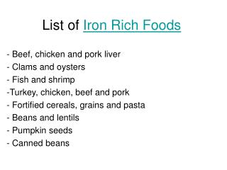
Iron metabolism (II) • Iron is mainly absorbed from the duedonum and proximal jejunum • EDTA, tannates, carbonates, oxaletes, phosphates, antiacites (magnesium trisilikat), clay decrease iron absorbtion • Ascorbic acid, citric acid, amino acides, sugars, gastric secretion and hydrochloric acid increase iron absorbtion • Normal diet contains 10-20 mg iron • Iron absorption is increased in the presence of iron deficiency and it decreases when there is iron overload
Iron metabolism (III) • Separate pathways exist for the absorption of heme and inorganic iron • The mechanism by which iron passes across the intestinal mucosa into the plasma has not yet been completely understood • The process appears to involve a ferrireductase, an integrin, a divalent iron transporter DMT-1, hephaestin, and ferroportin-1 • The main regulator of iron homeostasis seems to be the antimicrobial peptide hepcidin
Iron metabolism (IV) • Once ferric iron enters the plasma, it is bound by transferrin (iron binding protein) • Mono or divalent transferrin iron complex binds to transferrin receptor • The transferrin receptor is internalized together with bound transferrin and iron, and the iron is released inside the cell into an acidified vacuole • The transferrin receptor then moves back to the cell surface • Iron deficiency has adverse effects on activity of numerous enzymes, and in infants can result in impairment of growth and intellectual development. • The hematologic features of iron deficiency are non-specific and too often confused with other causes of microcytic anemia such as thalassemias, chronic disease, renal neoplasms, and other disorders.
Iron metabolism (V) • A low serum ferritin concentration is a good indicator of iron deficiency, but is elevated by inflammation, which lessens its sensitivity for the detection of iron deficiency coexisting with the anemia of chronic disease • The plasma iron is decreased and the iron binding capacity increased in severe iron deficiency, but these alterations are not uniformly present in mild iron deficiency • Low plasma iron levels are also characteristic of the anemia of chronic disease • Erythrocyte zinc protoporphyrin assay is a useful screening test but lacks specificity • Other laboratory tests that are useful include assays for serum transferrin receptor, erythrocyte ferritin concentration, and reticulocyte hemoglobin content
Iron deficiency (I) • Causes of iron deficiency • chronic blood loss • inadequate dietary iron intake • malabsorption of iron • diversion of iron to fetal and infant erythropoiesis during pregnancy and lactation • intravascular hemolysis with hemoglobinuria • combination of these factors • In men and in postmenopausal women. iron deficiency is most commonly caused by chronic bleeding from the gastrointestinal tract. • In the adult the most common causes of gastrointestinal bleeding are peptic ulcer, hiatal hernia, gastritis (including that due to alcohol or aspirin ingestion), hemorrhoids, vascular anomalies (such as angiodysplasia), and neoplasms
Iron deficiency (II) • Recurrent hemoptysis may lead to iron-deficiency anemia. It may be caused by congenital anomalies of the respiratory tract, endobronchial vascular anomalies, chronic infections, neoplasms, or valvular heart disease • Severe iron-deficiency anemia is a manifestation of idiopathic pulmonary hemosiderosis and Goodpasture syndrome • Menstrual bleeding is a very common cause of iron deficiency. The average menstrual blood loss is about 40 ml per cycle. Blood loss exceeds 80 ml (equivalent to about 30 mg of iron) per cycle in only 10 percent of women. The amount of menstrual blood lost does not seem to vary markedly from one cycle to another for any given individual
Iron deficiency (III) • Oral contraceptives reduce menstrual blood loss, but the use of an intrauterine coil for contraception increases menstrual blood loss, especially during the first year of use • Excessive bleeding may be caused by uterine fibroids and malignant neoplasms • Neoplasms, stones, or inflammatory disease of the kidney, ureter, or bladder may cause enough chronic blood loss to produce iron deficiency
Symptoms & Signs related with iron deficiency (I) • In younger patients with mild anemia no symptoms related with anemia is detected. But in the elderly there may be some symptoms of hypoxia even mild decrease in Hb level • In severe iron-deficiency anemia tachycardia with palpitations and pounding in the ears, headache, light-headedness, and even angina pectoris may occur • In the laryngopharynx, mucosal atrophy may lead to web formation in the postcricoid region, thereby giving rise to dysphagia (Paterson-Kelly/Plummer-Vinson syndrome) • Pica (the craving to eat unusual substances, such as dirt, clay, ice, laundry starch, salt, cardboard, or hair), is a classic manifestation of iron deficiency
Symptoms & Signs related with iron deficiency (II) • Hair loss may be a consequence of iron deficiency • In infants, iron deficiency is associated with poor attention span, poor response to sensory stimuli, and retarded behavioral and developmental achievement, even in the absence of anemia • The physical findings in iron-deficiency anemia include pallor, glossitis (smooth, red tongue), stomatitis, and angular cheilitis • Koilonychia, once a common finding, is now encountered rarely • Retinal hemorrhages and exudates may be seen in severely anemic patients (e.g., hemoglobin concentration 5 g/dl) • Splenomegaly is a rare finding
Iron deficiency anemia (Laboratory findings) • In severe uncomplicated iron-deficiency anemia; • the erythrocytes are hypochromic and microcytic • the plasma iron concentration is diminished • the iron-binding capacity increased • the serum ferritin concentration is low • the serum transferrin receptor is increased • erythrocyte zinc protoporphyrin concentration is increased • the marrow is depleted of stainable iron • Unfortunately, the classic combination of laboratory findings occurs consistently only when iron-deficiency anemia is far advanced, when there are no complicating factors such as infection or malignant neoplasms, and when there has not been previous therapy with transfusions or parenteral iron
Differential diagnoses of iron deficiency anemia • Thalassemias • Erythrocyte counts are decreased in iron deficiency anemia but normal or increased in thalassemia minor • RDW is increased in iron deficiency due to anisocytosis but normal in thalassemia minor • Hb A2 is increased in beta thalassemia minor (but should be tested after treatment of iron deficiency if present) • Plasma iron is normal or even increased and iron binding capacity is normal so, transferrin saturation is normal in thalassemia minor patients • Anemia of chronic diseases • In iron-deficiency anemia, the transferrin saturation is usually less than 16 percent, whereas in chronic diseases it is usually greater than 16 percent • Transferrin saturation is low but sometimes may be normal in iron-deficiency anemia, and conversely, low saturation is sometimes observed in chronic disease • Circulating transferrin receptors increase in iron deficiency but not in the anemia of chronic disease • The serum ferritin level is usually diminished in iron deficiency but it is generally increased in chronic inflammatory and neoplastic disorders • Stainable iron in the marrow is greatly decreased in amount or absent in iron deficiency anemia and normal or increased in the other disorders • Sideroblastic anemia • Reticulocytosis is usually absent • The serum iron and serum ferritin levels are generally normal or increased • Marrow examination shows increased amounts of iron
Treatment of iron deficiency anemia (I) • The reason of iron deficiency should be searched • Oral or parenteral iron should be administered (oral route is prefered) • Transfusion of RBCs is not recommended except emergency conditions • 100-200 mg/day elemantary iron should be taken orally in three or four doses 1 hour before meals • The first response to treatment is an increase of reticulocytes in circulation after 7-10 days • Treatment should continue to fill the iron stores after correcting anemia • If a response to treatment is not achieved it may be due to; • Patient’s non-complience to treatment • Absorption defect • Continuing blood loss • Incorrect diagnoses
Treatment of iron deficiency anemia (II) • Parenteral iron treatment is the following indications • malabsorption • intolerance to iron taken orally • iron need in excess of an amount that can be taken orally • non-compliance of the patient • Parenteral iron administration, together with erythropoietin, appears to alleviate the anemia that otherwise may complicate long-term dialysis treatment of patients with chronic renal disease • Iron sucrose (IV) or iron dextran (IM) is used • Several formulas for calculating the total iron dose for parenteral therapy have been proposed. • One example is as follows: Dose (mg iron) = [15 – patient’s hemoglobin (g/dL)] X body weight (kg) X 3