Download
1 / 26
260 likes | 398 Views
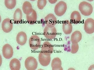
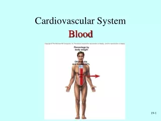
The Cardiovascular System: The Blood . Fluids of the Body . Cells of the body are serviced by 2 fluids blood composed of plasma and a variety of cells transports nutrients and wastes interstitial fluid bathes the cells of the body

E N D
Fluids of the Body • Cells of the body are serviced by 2 fluids • blood • composed of plasma and a variety of cells • transports nutrients and wastes • interstitial fluid • bathes the cells of the body • Nutrients and oxygen diffuse from the blood into the interstitial fluid & then into the cells • Wastes move in the reverse direction • Hematology is study of blood and blood disorders
Functions of Blood • Transportation • O2, CO2, metabolic wastes, nutrients, heat & hormones • Regulation • helps regulate pH through buffers • helps regulate body temperature • coolant properties of water • vasodilatation of surface vessels dump heat • helps regulate water content of cells by interactions with dissolved ions and proteins • Protection from disease & loss of blood
Physical Characteristics of Blood • Thicker (more viscous) than water and flows more slowly than water • Temperature of 100.4 degrees F • pH 7.4 (7.35-7.45) • 8 % of total body weight • Blood volume • 5 to 6 liters in average male • 4 to 5 liters in average female • hormonal negative feedback systems maintain constant blood volume and osmotic pressure
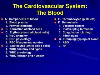
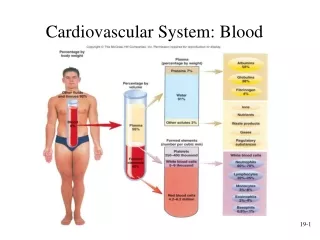
Components of Blood • Hematocrit • 55% plasma • 45% cells • 99% RBCs • < 1% WBCs and platelets
Blood Plasma • 0ver 90% water • 7% plasma proteins • created in liver • confined to bloodstream • albumin • maintain blood osmotic pressure • globulins (immunoglobulins) • antibodies bind to foreignsubstances called antigens • form antigen-antibody complexes • fibrinogen • for clotting • 2% other substances • electrolytes, nutrients, hormones, gases, waste products
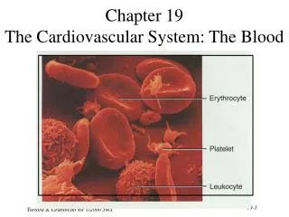
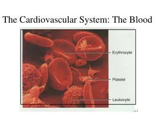
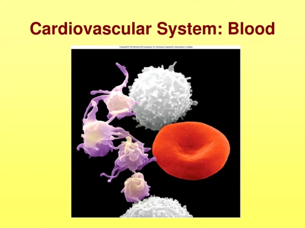
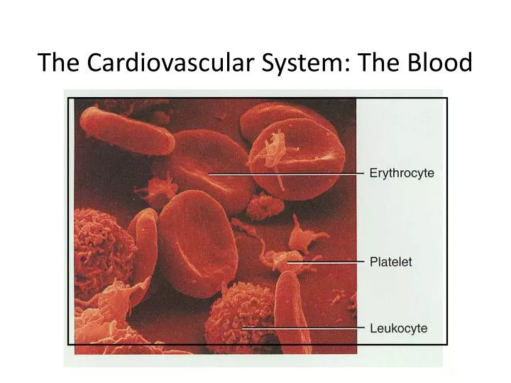
Formed Elements of Blood • Red blood cells ( erythrocytes ) • White blood cells ( leukocytes ) • granular leukocytes • neutrophils • eosinophils • basophils • agranular leukocytes • lymphocytes = T cells, B cells, and natural killer cells • monocytes • Platelets (special cell fragments)
Hematocrit • Percentage of blood occupied by cells • female normal range • 38 - 46% (average of 42%) • male normal range • 40 - 54% (average of 46%) • testosterone • Anemia • not enough RBCs or not enough hemoglobin • Polycythemia • too many RBCs (over 65%) • dehydration, tissue hypoxia, blood doping in athletes
Red Blood Cells or Erythrocytes • Contain oxygen-carrying protein hemoglobin that gives blood its red color • 1/3 of cell’s weight is hemoglobin • Biconcave disk 8 microns in diameter • increased surface area/volume ratio • flexible shape for narrow passages • no nucleus or other organelles • no cell division or mitochondrial ATP formation • Normal RBC count • male 5.4 million/drop ---- female 4.8 million/drop • new RBCs enter circulation at 2 million/second
Hemoglobin • Globin protein consisting of 4 polypeptide chains • One heme pigment attached to each polypeptide chain • each heme contains an iron ion (Fe+2) that can combine reversibly with one oxygen molecule
RBC Life Cycle • RBCs live only 120 days • wear out from bending to fit through capillaries • no repair possible due to lack of organelles • Worn out cells removed by fixed macrophages in spleen & liver • Breakdown products are recycled
Erythropoiesis: Production of RBCs • Proerythroblast starts to produce hemoglobin • Many steps later, nucleus is ejected & a reticulocyte is formed • orange in color with traces of visible rough ER • Reticulocytes escape from bone marrow into the blood • In 1-2 days, they eject the remaining organelles to become a mature RBC
WBC Anatomy and Types • All WBCs (leukocytes) have a nucleus and no hemoglobin • Granular or agranular classification based on presence of cytoplasmic granules made visible by staining • granulocytes are neutrophils, eosinophils or basophils • agranulocytes are monocyes or lymphocytes
WBC Physiology • Less numerous than RBCs • 5000 to 10,000 cells per drop of blood • 1 WBC for every 700 RBC • Leukocytosis is a high white blood cell count • microbes, strenuous exercise, anesthesia or surgery • Leukopenia is low white blood cell count • radiation, shock or chemotherapy • Only 2% of total WBC population is in circulating blood at any given time • rest is in lymphatic fluid, skin, lungs, lymph nodes & spleen
Differential WBC Count • Detection of changes in numbers of circulating WBCs (percentages of each type) • indicates infection, poisoning, leukemia, chemotherapy, parasites or allergy reaction • Normal WBC counts • neutrophils 60-70% (up if bacterial infection) • lymphocyte 20-25% (up if viral infection) • monocytes 3 -- 8 % (up if fungal/viral infection) • eosinophil 2 -- 4 % (up if parasite or allergy reaction) • basophil <1% (up if allergy reaction or hypothyroid)
Bone Marrow Transplant • Intravenous transfer of healthy bone marrow • Procedure • destroy sick bone marrow with radiation & chemotherapy • donor matches surface antigens on WBC • put sample of donor marrow into patient's vein for reseeding of bone marrow • success depends on histocompatibility of donor & recipient • Treatment for leukemia, sickle-cell, breast, ovarian or testicular cancer, lymphoma or aplastic anemia
Platelets--Life History • Platelets form in bone marrow by following steps: • myeloid stem cells to megakaryocyte-colony forming cells to megakaryoblast to megakaryocytes whose cell fragments form platelets • Short life span (5 to 9 days in bloodstream) • formed in bone marrow • few days in circulating blood • aged ones removed by fixed macrophages in liver and spleen
Complete Blood Count • Screens for anemia and infection • Total RBC, WBC & platelet counts; differential WBC; hematocrit and hemoglobin measurements • Normal hemoglobin range • infants have 14 to 20 g/100mL of blood • adult females have 12 to 16 g/100mL of blood • adult males have 13.5 to 18g/100mL of blood
Hemostasis • Stoppage of bleeding in a quick & localized fashion when blood vessels are damaged • Prevents hemorrhage (loss of a large amount of blood) • Methods utilized • vascular spasm • platelet plug formation • blood clotting (coagulation = formation of fibrin threads)
Blood Clotting • Blood drawn from the body thickens into a gel • gel separates into liquid (serum) and a clot of insoluble fibers (fibrin) in which the cells are trapped • If clotting occurs in an unbroken vessel is called a thrombosis • Substances required for clotting are Ca+2, enzymes synthesized by liver cells and substances released by platelets or damaged tissues • Clotting is a cascade of reactions in which each clotting factor activates the next in a fixed sequence resulting in the formation of fibrin threads • prothrombinase & Ca+2 convert prothrombin into thrombin • thrombin converts fibrinogen into fibrin threads
Overview of the Clotting Cascade • Prothrombinase is formed by either the intrinsic or extrinsic pathway • Final common pathway produces fibrin threads
Clot Retraction & Blood Vessel Repair • Clot plugs ruptured area of blood vessel • Platelets pull on fibrin threads causing clot retraction • trapped platelets release factor XIII stabilizing the fibrin threads • Edges of damaged vessel are pulled together • Fibroblasts & endothelial cells repair the blood vessel
Anemia = Not Enough RBCs • Symptoms • oxygen-carrying capacity of blood is reduced • fatigue, cold intolerance & paleness • lack of O2 for ATP & heat production • Types of anemia • iron-deficiency =lack of absorption or loss of iron • pernicious = lack of intrinsic factor for B12 absorption • hemorrhagic = loss of RBCs due to bleeding (ulcer) • hemolytic = defects in cell membranes cause rupture • thalassemia = hereditary deficiency of hemoglobin • aplastic = destruction of bone marrow (radiation/toxins)
Sickle-cell Anemia (SCA) • Genetic defect in hemoglobin molecule (Hb-S) that changes 2 amino acids • at low very O2 levels, RBC is deformed by changes in hemoglobin molecule within the RBC • sickle-shaped cells rupture easily = causing anemia & clots • Found among populations in malaria belt • Mediterranean Europe, sub-Saharan Africa & Asia • Person with only one sickle cell gene • increased resistance to malaria because RBC membranes leak K+ & lowered levels of K+ kill the parasite infecting the red blood cells
Hemophilia • Inherited deficiency of clotting factors • bleeding spontaneously or after minor trauma • subcutaneous & intramuscular hemorrhaging • nosebleeds, blood in urine, articular bleeding & pain • Hemophilia A lacks factor VIII (males only) • most common • Hemophilia B lacks factor IX (males only) • Hemophilia C (males & females) • less severe because alternate clotting activator exists • Treatment is transfusions of fresh plasma or concentrates of the missing clotting factor
Leukemia • Acute leukemia • uncontrolled production of immature leukocytes • crowding out of normal red bone marrow cells by production of immature WBC • prevents production of RBC & platelets • Chronic leukemia • accumulation of mature WBC in bloodstream because they do not die • classified by type of WBC that is predominant---monocytic, lymphocytic.