Download
1 / 27
270 likes | 387 Views
Blood and the Cardiovascular System. By Arielle Haddad, Jackie Lazo , Jackie Mendoza, and Lauren Walsh. Components of Blood. Blood: the only fluid tissue; complex connective tissue Formed elements: living blood cells Plasma : nonliving fluid matrix

E N D
Blood and the Cardiovascular System By Arielle Haddad, Jackie Lazo, Jackie Mendoza, and Lauren Walsh
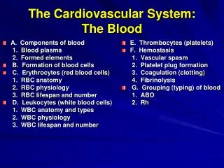
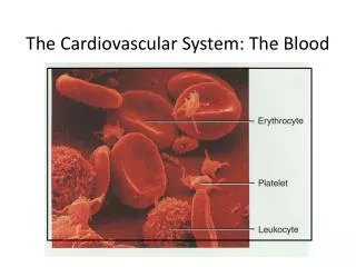
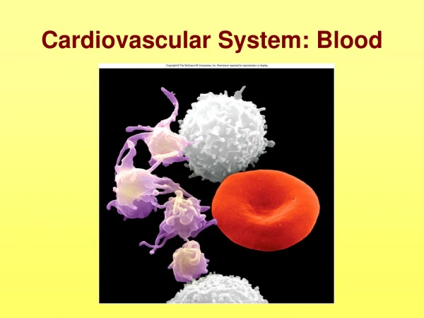
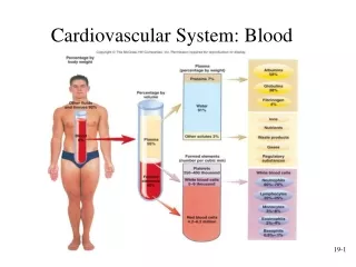
Components of Blood • Blood: the only fluid tissue; complex connective tissue • Formed elements: living blood cells • Plasma: nonliving fluid matrix • Erythrocytes: red blood cells- oxygen transport • Buffy coat: in between formed elements and plasma- location of leukocytes and platelets (blood clotting) • Heavier and 5 times thicker than water
Plasma • 90% water • Includes nutrients, electrolytes, respiratory gases, hormones, proteins, and waste • Plasma proteins: keep water in bloodstream, clot blood, protect body from pathogens • Plasma helps to distribute body heat evenly.
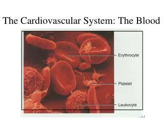
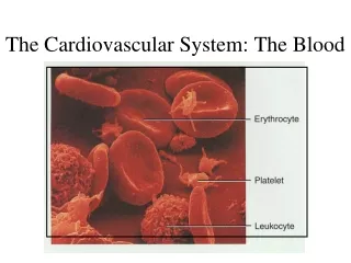
Formed Elements: Erythrocytes • Red blood cells • Deliver oxygen to all cells in the body • Anucleate: lack a nucleus • Flattened disc shape • Contain hemoglobin: iron-bearing protein which transports the oxygen • Outnumber white blood cells 1000 to 1
Formed Elements: Leukocytes • White blood cells • Defend body against bacteria, viruses, parasites, and tumor cells • Have the ability to enter and exit the bloodstream- diapedesis • Can locate infections and damage by responding to chemicals diffused by damaged tissue- positive chemotaxis • Two major groups: Granulocytes and Agranulocytes • Granulocytes: acute infections, allergies, parasites • Agranulocytes: immune response, chronic infections
Formed Elements: Platelets • Fragments of multinucleate cells- megakaryocytes • Pieces quickly seal themselves off from surrounding fluids
Hemostasis • If a blood vessel wall breaks, series of actions are set in motion to accomplish hemostasis. • Hemostasis– stoppage of blood flow • Three major stages: • Platelet plug formation • Vascular spasms • Coagulation (blood clotting)
Stages of Hemostasis • Platelets plug forms - platelets become sticky and cling to the damaged site; release chemicals to attract more platelets to site • Vascular spasms occur- platelets release serotoninwhich causes vessel to go into spasms; decreases blood loss until clotting • Coagulation events occur • Tissue factor (TF) is released; • PF3, a phospholipid, • Interacts with TF and triggers clotting cascade.; • Prothrombin activator converts prothrombin into thrombin; • Thrombin joins fibrinogen proteins into long hairlike molecules of insoluble fibrin, trapping red blood cells and creates clot; • Clot reacts and retracts, squeezing serum (plasma minus clotting proteins) from mass and bringing the blood vessel closer together
Disorders of Hemostasis • Undesirable Clotting • Thrombus- clot that develops and persists in an unbroken blood vessel • Embolus- a thrombus that has broken away from vessel and into bloodstream • Bleeding Disorders • Thrombocytopenia- the presence of relatively few platelets in blood • Hemophilia- inherited clotting defect caused by absence of blood-clotting factor
Blood Groups and Transfusions • Human Blood Groups • Antigen- substance the body recognizes as foreign • Antigens stimulate the body to release antibodies to mount defense against it • Antibody- specialized substance produced by body to provide immunity against antigens • Agglutination- the binding of antibodies that causes RBCs to clump and causes clotting
Blood Groups and Transfusions • ABO blood groups are based on which two antigens, type A or type B, a person inherits • Absence of both antigens = type O blood • Presence of both antigens= type AB blood • Rhesus (Rh) blood group refers to the 5 main Rhesus antigens (C, c, D, E and e) as well as the many other less frequent Rhesus antigens • Hemolysis- rupture of RBCs
Blood Vessels Arteries Veins Capillaries
What are blood vessels? • Blood vessels are intricate networks of hollow tubes that transport blood throughout the entire body. • 3 types of blood vessels: • Arteries • Veins • Capillaries
Arteries • The heart pumps blood out through one main artery called the dorsal aorta. • The main artery then divides and branches out into many smaller arteries so that each region of your body has its own system of arteries supplying it with fresh, oxygen-rich blood. • The smallest arteries are called arterioles which branch into capillaries. • Arteries are tough on the outside and smooth on the inside. • An artery actually has three layers: an outer layer of tissue, a muscular middle, and an inner layer of epithelial cells. • The muscular wall of the artery helps the heart pump the blood. • When the heart beats, the artery expands as it fills with blood. • When the heart relaxes, the artery contracts, exerting a force that is strong enough to push the blood along.
Structure of Arteries • The artery wall consists of three layers: • Tunica Adventitia • Tunica Media • Tunica Intima • Tunica Adventitia: The tunica adventitia is the strong outer covering of arteries and veins. It is composed of connective tissue as well as collagen and elastic fibers. These fibers allow the arteries and veins to stretch to prevent overexpansion due to the pressure that is exerted on the walls by blood flow.
Structure of Arteries (cont.) • Tunica Media: The tunica media is the middle layer of the walls of arteries and veins. It is composed of smooth muscle and elastic fibers. This layer is thicker in arteries than in veins. • Tunica Intima: The tunica intima is the inner layer of arteries and veins. In arteries this layer is composed of an elastic membrane lining and smooth endothelium that is covered by elastic tissues.
Veins • Veins are similar to arteries but, because they transport blood at a lower pressure, they are not as strong as arteries. • Like arteries, veins have three layers: • an outer layer of tissue • muscle in the middle • a smooth inner layer of epithelial cells. Veins receive blood from the capillaries after the exchange of oxygen and carbon dioxide has taken place. • the veins transport waste-rich blood back to the lungs and heart. • It is important that the waste-rich blood keeps moving in the proper direction and not be allowed to flow backward. This is accomplished by valves that are located inside the veins. • The valves are like gates that only allow traffic to move in one direction.
Veins (cont) • The walls of the veins are rather thin, the waste-rich blood is visible through the skin on some parts of the body. • For examle: • Wrist • Hands • Ankles • Overview: • carry blood away from the heart • carry blood to the heart • divide into two bronchial tubes • are controlled by the cerebrum
Capillaries • Capillaries are so small that red blood cells can only travel through them in single file. • Capillaries are extremely small vessels located within the tissues of the body that transport blood from the arteries to the veins. • Capillary walls are thin and are composed of endothelium (a single layer of overlapping flat cells). • Oxygen, carbon dioxide, nutrients and wastes are exchanged through the thin walls of the capillaries. • The flow of blood is controlled by structures called precapillary sphincters. • These structures are located between arterioles and capillaries and contain muscle fibers that allow them to contract. • When the sphincters are open, blood flows freely to the capillary beds of body tissue. • When the sphincters are closed, blood is not allowed to flow through the capillary beds.
The Heart • 4 chambers ( or compartments ) • 2 upper chambers : Left Atrium Right Atrium • 2 lower chambers : Left Ventricle Right Ventricle
Lub Dub If you listen to your heartbeat, it makes a lub dub sound. The lub is when blood is pushed out of the heart into the body and the dub is the reloading of the heart with more blood ready to push it out to the body.
Blood Flow through the Human Body Blood with little oxygen is pumped from the Right Atrium through a valve to the Right Ventricle and through another valve into the Pulmonary Vein. The Pulmonary Vein sends it to the Lungs to pick up Oxygen. It’s sent from the Lungs through Pulmonary Arteries to the Left Atrium through a valve to the Left Ventricle. The Left Ventricle sends it through a valve to the Aorta. The Aorta is a series of branches that sends oxygenated blood throughout the body.
Heart Facts • Hold out your hand and make a fist. If you're a kid, your heart is about the same size as your fist, and if you're an adult, it's about the same size as two fists. • Your heart beats about 100,000 times in one day and about 35 million times in a year. During an average lifetime, the human heart will beat more than 2.5 billion times. • Give a tennis ball a good, hard squeeze. You're using about the same amount of force your heart uses to pump blood out to the body. Even at rest, the muscles of the heart work hard--twice as hard as the leg muscles of a person sprinting.