Download
1 / 58
580 likes | 707 Views
Cardiovascular System: The Blood. Dr. Michael P. Gillespie. Constituents Of Blood. Blood is a connective tissue composed of a liquid matrix called plasma that dissolves and suspends various cells and fragments. Interstitial fluid is the fluid that bathes body cells. Functions of Blood.

E N D
Cardiovascular System: The Blood Dr. Michael P. Gillespie
Constituents Of Blood • Blood is a connective tissue composed of a liquid matrix called plasma that dissolves and suspends various cells and fragments. • Interstitial fluid is the fluid that bathes body cells.
Functions of Blood • Transportation – oxygen, carbon dioxide, nutrients, hormones, heat, & waste products. • Regulation – maintains homeostasis (ph, heat, osmotic pressure). • Protection – clotting, WBCs, & antibodies.
Physical Characteristics Of Blood • Blood is denser and more viscous than water. • The temperature is 38 degrees C (100.4 degrees F). • Slightly alkaline ph ranging from 7.35 to 7.45. • 20% of the extracellular fluid (about 8% of total body mass). • The blood volume is 5 to 6 liters (1.5 gal) in an average adult male and 4 to 5 liters (1.2 gal) in an average adult female.
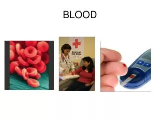
Withdrawing Blood • Blood samples for laboratory testing are obtained in various ways. • Venipuncture (the most common method). • A finger or heel stick is utilized for children and people who must monitor their blood daily (I.E. Diabetics). • An arterial stick is utilized when the level of O2 must be determined.
Components Of Blood • Whole blood has two components: • Blood plasma, a watery liquid matrix that contains dissolved substances. • Formed elements (cells and cell fragments). • 45% formed elements and 55% plasma.
Blood Plasma • A straw-colored liquid which is about 91.5% water and 8.5% solutes. • Plasma proteins – maintain osmotic pressure. • Albumins, globulins, and fibrinogen (synthesized by hepatocytes). • Gamma globulins (antibodies 0r immunoglobulins). • Electrolytes, nutrients, regulatory substances (i.E. Enzymes and hormones), gases, & waste products (urea, uric acid, creatinine, ammonia, and bilirubin).
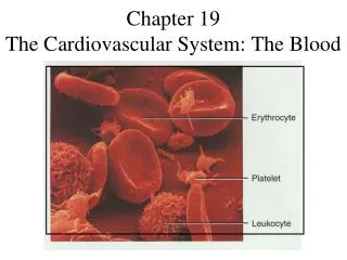
Formed Elements • RBCs. • WBCs – perform many functions.
Formed Elements • Platelets – cell fragments.
Formed Elements • Hematocrit – the percentage of total blood occupied by RBCs. • Males have a higher hematocrit than females because testosterone stimulates the production of erythropoeitin (EPO). • Menstruation leads to lower values for females during their reproductive years. • Anemia is a significant drop in the hematocrit. • Polycythemia is an. • Abnormally high percentage of RBCs.
Causes of Polycythemia • Abnormal increase in RBC production. • Tissue hypoxia. • Dehydration. • Blood doping or use of Epoetin alfa (Procrit or Epogen) by athletes. • This increases the work load of the heart. • The increased #s of RBCs raise the viscosity of the blood, which increases the resistance to blood flow. This can cause high blood pressure and stroke.
Formation of Blood Cells • Hemopoiesis (hematopoiesis) – the process by which the formed elements of blood develop. • Blood cells, macrophages, reticular cells, mast cells, and adipocytes arise from the red bone marrow. • Pluripotent stem cells in the bone marrow reproduce themselves, proliferate and differentiate into mature blood cells.
Two Types of Pluripotent Stem Cells • Myeloid stem cells. • Give rise to red blood cells, platelets, monocytes, neutrophils, eosinophils, and basophils. • Lymphoid stem cells. • Give rise to lymphocytes.
Generations Of Cell Lines In The Development Of Blood Cells • Pluripotent stem cells – mesenchymal cells which have the capacity to develop into many different types of cells. They can reproduce themselves. • Progenitor cells – cannot reproduce themselves. • Precursor cells (blasts) – they develop into the actual formed elements of the blood.
Hormones That Regulate Blood Cell Development • Hemopoietic growth factors – regulates differentiation and growth of progenitor cells. • Erythropoietin (EPO) from the kidneys – increases the # of RBC precursors. • Thrombopoietin (TPO) from the liver – stimulates the formation of platelets. • Colony-stimulating factors (CSFs) or interleukins stimulate WBC formation.
Medical Uses of Hemopoietic Growth Factors • EPO is utilized in end stage kidney disease to increase RBC formation. • CSFs are utilized to stimulate WBC formation in cancer patients undergoing chemotherapy. • Thrombopoietin helps induce platelet formation in chemotherapy patients.
Red Blood Cells • Red blood cells (RBCs) or erythrocytes: • Contain hemoglobin – oxygen-carrying protein which gives the cell its color. • Approximately 5 million RBCs are present per microliter of blood. • Approximately 2 million RBCs are created and destroyed per second.
RBC Anatomy • Biconcave discs with a diameter of 7-8 micrometers. • The plasma membrane is flexible, which allows them to deform without rupturing as they squeeze through capillaries. • RBCs lack a nucleus and other organelles. • RBCs cannot reproduce or carry on extensive metabolic activities.
RBC Physiology • With no nucleus, RBCs have more space available for oxygen transport. • RBCs lack mitochondria and generate ATP anaerobically; Consequently, they do not use up the oxygen they are transporting.
RBC Physiology • The biconcave disc has a greater surface area allowing greater diffusion of gas molecules. • Hemoglobin binds to oxygen, carbon dioxide, and nitric oxide (NO). • NO causes vasodilation which enhances oxygen delivery to cells.
RBC Life Cycle • Red blood cells live only about 120 days. • Macrophages in the spleen and liver remove dead RBCs through phagocytosis. • Hemoglobin is broken down into its globin and heme portions. • Globin is broken down into amino acids, which are reused for proteins. • Heme is converted into the yellow-orange pigment bilirubin.
Reticulocyte Count • The rate of eryhtropoiesis is measured by a reticulocyte count. • Low “retic” count – could indicate a shortage of erythropoietin due to a nutritional deficiency or leukemia. • High “retic” count – could indicate a good response to previous blood loss, iron therapy, or illegal use of Epoetin by an athlete.
White Blood Cells • White blood cells (WBCs) or leukocytes have a nucleus and do not contain hemoglobin. • Classified as either granular or agranular.
Types Of White Blood Cells • Eosinophil • Basophil • Neutrophil • Small lymphocyte • Monocyte
Numbers Of WBCs • RBCs outnumber WBCs by about 700:1. • There are approximately 5000 – 10,000 cells per microliter of blood. • Leukocytosis – an increase in the number of WBCs due to stresses such as microbes, strenuous exercise, anesthesia, or surgery. • Leukopenia – a decrease in the number of WBCs due to radiation, shock, or chemotherapy.
Functions Of WBCs • The WBCs combat pathogens by phagocytosis and other immune responses. • WBCs leave the bloodstream by emigration.
Functions Of WBCs • Neutrophils and macrophages are active in phagocytosis. • Phagocytes are attracted to inflamed tissues through a process called chemotaxis. • Phagocytes release the enzyme lysozyme, which destroys certain bacteria.
Functions Of WBCs • Eosinophil – release histamines. Respond to allergic responses and parasitic infection. • Basophil – liberate heparin, histamine, and serotonin. They intensify the inflammatory reaction and are involved in hypersensitivity (allergic) reactions.
Functions Of WBCs • Neutrophil – active in phagocytosis and ingest bacteria and dead matter. They respond to bacteria and fungi. • Lymphocyte – “soldiers” of the immune system. • Monocyte – turn into macrophages and clean up cellular debris after an infection.
Differential WBC Count • Utilized to detect infection, inflammation, poisoning, blood disorders, effects of chemotherapy, allergic reactions, and parasitic infections.
Platelets • The hormone thrombopoietin influences the production of platelets (thrombocytes). • Platelets help stop blood loss by forming a platelet plug.
Complete Blood Count (CBC) • A test that screens for anemia and various infections. • Counts of RBCs, WBCs, platelets, hematocrit, and a differential white blood cell count are included.
Blood Clotting • Serum is a straw colored liquid and the gel is called a clot. • The process of gel formation is called clotting or coagulation. • Clotting factors are involved in the coagulation cascade.
Blood Clotting • Normal clotting requires vitamin K, which is produced by bacteria in the intestines. • Dissolution of a clot is called fibrinolysis. • Anticoagulants (heparin & Warfaring a.K.A. Coumadin) are utilized for patients at risk of forming a blood clot.
Intravascular Clotting • Thrombosis – clotting in an unbroken blood vessel. • Thrombus – the clot itself. • Embolus – a blood clot, bubble of air, fat from broken bones, or a piece of debris transported by the bloodstream. • Pulmonary embolism – when an embolus lodges in the lungs.