Download
1 / 46
480 likes | 713 Views
Antigen presentation in CNS. Basic antigen presentation. Antigen is in context of MHC Antigen presenting cells (APC) MHC I interacts with CD8 T cells MHC II interacts with CD4 T cells. APCs outside of brain. Professional Dendritic cells B cells Macrophages Target APCs

E N D
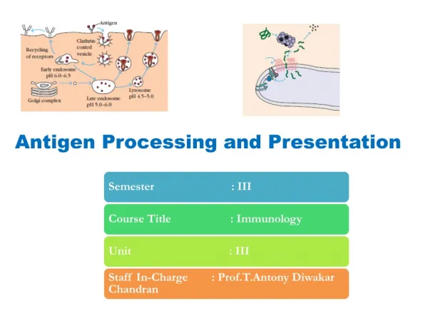
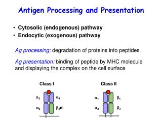
Basic antigen presentation • Antigen is in context of MHC • Antigen presenting cells (APC) • MHC I interacts with CD8 T cells • MHC II interacts with CD4 T cells
APCs outside of brain • Professional • Dendritic cells • B cells • Macrophages • Target APCs • Most cells of body
Fig 1.4 part 1 of 2
Roles of APC • MHC II+ APC initiate immune response by activating either CD4 or CD8 T cells • Professional APC activating CD8 T cell is called cross-priming • MHC I allows infected cells to be killed by CTL
Costimulation • Costimulation of T cell by second signal required for activation • MHC/TCR interaction without costimulation results in anergy • B7.1 and B7.2 interact with CD28 on T cell • CD40/CD40L is another costimulatory pair • CTLA-4 on T cell is negative signal
CD4 and CD8 • CD4 T cells are helper T cells • Th1 generally promote cell mediated responses and nonspecific inflammation • Th2 generally promote humoral (B cell mediated) immunity • CD8 T cells are usually Cytotoxic T cells (CTL)
APC ICAM-1 LFA-3 MHC T cell LFA-1 CD2 CD4 and CD8 Adhesion also important for activation of T cells
Cell Types in the Nervous System • Neurons • Glia • Endothelial cells (blood vessels) Photos from Burkitt Functional Histology
Glia • Oligodendrocytes (myelin) • Astrocytes (or astroglia) • Ependymal cells (and choroid plexus) • Microglia
Endothelial Cells • Form Blood Brain Barrier
Microglia Bacskai 2001
Historical Bkgd of Immune Privilege • Antigen presentation necessary for graft rejection • Skin grafts implanted in brain survive longer than other places in body • Concept of immunologically privileged site (Medawar, 1948)
Immune Privilege • Earlier studies done w tumors implanted in brain in 1920s • Medawar did skin grafts • Graft to non-immunized rabbit not rejected • Graft to pre-immunized rabbit rejected • Implies afferent arm deficient
Afferent/Efferent • Afferent: presentation of antigen to generate specific T cell or B cell activation • Efferent: Effector mechanism capable of eliminating problem (eg, CTL)
Other privileged sites • Eye, anterior chamber • Testis • Placenta
Some antigen presentation does occur • After graft to brain, peripheral grafts are more efficiently rejected
CNS situations where Antigen presentation may occur • Autoimmunity • Neurodegenerative diseases • Viral infections
Candidate APCs in CNS • Professional • Microglia • Macrophages • Choroid Plexus Epithelial cells • Astrocytes • Oligodendrocytes • Perivascular cells • Endothelial cells • Smooth muscle/pericytes
Candidate APCs in CNS • Targets • Neurons • Oligodendrocytes • Astrocytes
Types of Microglia in CNS • Microglia in parenchyma • Perivascular macrophages • Macrophages in meninges and CP
Macrophages • Formed in bone marrow • Some circulate as monocytes • Some resident in tissue • Phagocytose • Can contribute to activation of T cells, but also respond to cytokines released by T cells • Microglia and macrophages share all markers
Activation of microglia • Normally quiescent • Activation changes shape • Shorter, more processes • Activation changes markers • MHC I, MHC II, CD4 • Two stages to activation • Shape and markers • Phagocytic activity • IFN and plasma proteins (damage)
Activated microglia • Peripheral macrophages attract neutrophils, increase inflammation • CNS response is delayed by days
Microglia turnover • Slow, continuous • Local cell division and invasion from blood (how HIV gets in) • Thymidine incorporation • Bone marrow transplant • GFP
Are microglia APCs? • Not before activation • After activation some evidence contribute as APC (regions of activated MG are more affected in EAE), but some evidence suppressive • IFN activated MG stimulate naïve CD4 T cells and Th1 T cells
Other brain macrophages as APC • Better evidence that macrophages of Choroid Plexus act as APC • Perivascular cells act as APC
Location of normal antigen presentation • Antigen travels to draining lymph nodes, some carried by APC • Less efficient drainage means less efficient presentation, less vigorous response • Implies MHC II not absolutely necessary in tissue • Antigen presentation by microglia to T cells initially stimulated in periphery
Astrocytes • 10x astrocytes as neurons • Astrocyte locations, clues to function • At interfaces (blood vessels, ependyma, pia) • Near neuron communication sites (Nodes of Ranvier, synapses) • Protoplasmic and Fibrous types • Proto in gray matter, short many processes (Type 1) • Fibrous in white matter, few but long processes (type 2)
Astrocyte Functions • Ion, water homeostasis • Neurotr. homeostasis • Also receptors for Nts • Provide precursors for Nts • Remove excitotoxic Nts • Cellular commun. (Ca waves) • Energy metab • Detox and injury responses • Neurotrophic • Immune Function
Astrocyte Injury responses • Proliferation, hypertrophy • Swelling (cranial pressure) • Release glutamate • GFAP increases • Profusion of processes (gliosis)
Astrocytes in immune disorders • MS: “sclerosis” is gliosis • EAE: reactive astrocytes • HIV: astrocytes may increase HIV replication in microglia
Produced by Astrocytes MHC I MHC II IL-1 TNF TGF CSF ICAM Complement C3fB Astrocyes respond to IL-1 IL-1 IFN TNF LPS TGF Immune Factors
Regional diffs in Astrocytes • Vary in abiltiy to take up glu and 5HT • vary in presence of Nt receptors • neurotrophic effects
Astrocytes as APC? • Only appear to function as APC in vitro, not in vivo • Nts appear to downregulate MHC responses • “In essence, therefore there is no evidence…” p 378