Download
1 / 55
670 likes | 1.11k Views
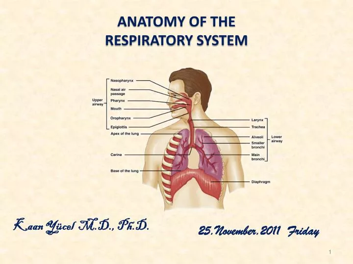
Anatomy of the resPIRATORY SYSTEM. Kaan Yücel M.D., Ph.D . 25.November.20 1 1 Friday. nose & assocIated structures . Nose is divisible into two parts as external nose and nasal cavity. Functions of the nose and the nasal cavities are: Olfaction (sense of smell) Respiration

E N D
Anatomy of the resPIRATORY SYSTEM Kaan Yücel M.D., Ph.D. 25.November.2011 Friday
nose & assocIated structures • Nose is divisible into two parts as external nose and nasal cavity. • Functions of the nose and the nasal cavities are: • Olfaction (sense of smell) • Respiration • Filtration of the dust in the inspired air • Humidification and warming of the inspired air • Reception of the secretions from the paranasal sinuses and nasolacrimal ducts
EXTERNAL NOSE • Extends the nasal cavities onto the front of the face and positions the nares so that they point downward. • Pyramidal in shape with its apex anterior in position.
External Nose has five parts: • Dorsum • Root • Apex • Nares (nostrils, anterior nasal apertures) • Alae of the nose • External nose has bony and cartilaginous parts.
Bones contributing to the structure of the external nose: • Nasal bones • Frontal process of maxilla • Nasal part of frontal bone
Cartilages contributing to the structure of the external nose: • Lateral cartilages (paired) • Alar cartilages (paired) • Septal cartilage (single)
Nasal Cavities: • The two nasal cavities are the uppermost parts of the respiratory tract. • They contain the olfactory receptors. • The nasal cavities are separated: • from each other by a midline nasal septum • from the oral cavity below by the hard palate • from the cranial cavity above by parts of the frontal, ethmoid, and sphenoid bones.
Nasal septum is composed of three structures: • Perpendicular plate of the ethmoid bone • Vomer • Septal cartilage
Eachnasalcavity is dividedintoolfactoryarea (upper 1/3) andrespiratoryarea (lower2/3). • Posteriorly, each nasal cavity communicates with the nasopharynx through two openings calledchoana.
Walls of the nasal cavity • Roof of the nasal cavity (anterior to posterior) • nasal bone • frontal bone • cribriform plate of the ethmoid bone • body of the sphenoid bone
Floor of the nasal cavityis formedbythehard palate (palatine process of maxilla and horizontal plate of the palatine bone). Atrium: Anteriorpart of middlenasalmeatus
Lateral wall of the nasal cavity (anterior to posterior) • frontal process of maxilla • lacrimal bone • superior nasal concha (of the ethmoid bone), middle nasal concha (of the ethmoid bone), inferior nasal concha • perpendicular plate of the palatine bone • medial lamina of the pterygoidprocess
Medial wall of the nasal cavity is formed by the nasal septum. Themedialwall has a smoothsurface, whereasthelateralwall is unevenduetotheexistance of thenasalconchae.
The spaces between the nasal conchae and the lateral wall of the nasal cavity are called the meatus. • Superior nasal meatus • Middle nasal meatus • Inferior nasal meatus
The following sinuses open into the middle nasal meatus • Frontalsinus • Maxillarysinus • Ethmoid air cells • Sphenoidsinusopensintothesphenoethmoidrecess • Nasolacrimal ductopensintotheinferior nasal meatus
Arterialsupply of thenose • The nose has an extensive arterial supply. The branches of the maxillary, ophthalmic and facial arteries supply the nose. • Veinsof thenose • There is a rich network of veinsdeeptothemucosa of thenose. • Thisvenous network is important in warmingtheairbefore it entersthetracheaandthelungs.
Nerves of thenose Sensory innervation of the nose is mainly from themaxillarynerve and the ophthalmic nerve.
Nerves of thenose Sensory innervation of the nose is mainly from themaxillarynerve and the ophthalmic nerve. Withintheepithelium of theolfactoryregionliestheolfactorycells (neurons). Theperipheralprocesses of thesecellsterminateunderthemucosaandaresensitivetoodourmolecules in theair. Thecentralprocessesformstheolfactorynerves (CN I).
Paranasalsinuses • Paranasal sinuses are air filled spaces lying within the bones around the nasal cavity. • The paranasal sinuses develop as outgrowths from the nasal cavities and erode into the surrounding bones. All are: • lined by respiratory mucosa, which is ciliated and mucus secreting; • open into the nasal cavities; and • innervated by branches of the trigeminal nerve [V].
Sinuses are named according to the bones they are located in: • Frontal sinuses • Ethmoid sinuses • Sphenoid sinuses • Maxillary sinuses
Frontal sinus • The frontal sinuses, one on each side, are variable in size and are the most superior of the sinuses. • Thefrontalsinuslies within the inner and outer plates of the frontal bone, posterior to the supercilliary arches and the root of the nose. • It drains into the middle nasal meatus.
Ethmoidsinuses • Several ethmoid air cells (3-15) collectively are called the ethmoid sinuses. • Ethmoid air cells form three groups: • Anterior group • Middle group • Posterior group
Sphenoid sinus • The sphenoid sinus is situated within the body of the sphenoid bone. • Sinusesof eachside is seperatedby a bonyseptum. • Itdrains into the sphenoethmoidal recess.
Maxillary sinus • The maxillary sinuses, one on each side, are the largest of the paranasal sinuses and completely fill the bodies of the maxillae. • The maxillary opening drains into the middle nasal meatus.
Larynx • Larnynxis the organ of phonation (vocalization). • It is formed of cartilage, muscles and connective tissue. • Larynx’sinnersurface is coveredbytherespiratorymucosa.
Thecavity of thelarynx iscontinuousbelowwiththetrachea, andaboveopensintothepharynx (nasopharynx).
Skeleton of larynxis formed of 3 unpaired and 3 paired cartilages • Unpaired cartilages • Thyroid cartilage (biggest) • Cricoid cartilage • Epiglottic cartilage • Paired cartilages • Arytenoid • Corniculate • Cuneiform
Thyroid cartilage • The thyroid cartilage is the largest cartilage of the larynx. • It is formed of two laminae which fuse anteriorly at the thyroid angleto form laryngeal prominence(Adam’s apple).
Fibroelasticmembrane of thelarynx • It lies under the mucosa of the larynx. • The fibroelastic membrane of the larynx has thickenings at certain regions and forms some of the ligaments between the cartilages. • It is formed of two parts: • Quadrangular membrane • Conus elasticus
Conuselasticus (cricovocal membrane):Itsfree upper margin thickens to form the vocal ligament, which is covered by mucosa to form the vocal fold. The opening between the two vocal folds is called rimaglottis. Vocalcord= Vocalfold
Eachvocal ligament, convergesanteriorly andattaches to the anterior part of the inner surface of the thyroid cartilage (thyroid angle). • Posteriorly, theyindividuallyattach to the vocal processes of the arytenoid cartilages.
Rimaglottis widens during inspiration and two vocal folds are approximated during phonation. • Various changes of the vocal folds determine the color, pitch and the tones of sound. • Pitch increases with tensing, decreasesbyrelaxation. • Intensity of expiration determines the loudness of sound.
Laryngeal Muscles • Extrisic muscles • Thesearethe suprahyoid and infrahyoid muscles. • Theyeither depress or elavate the larynx and hyoid bone. • Intrinsic muscles: There aresixintrinsicmuscles in thelarnyx. Theymovethe laryngeal parts.
Vasculature of the larynx Themajorbloodsupplytothelarynx is bythesuperiorandinferiorlaryngealarteries.
Nerves of the larynx Larynx is innervated by the inferiorandsuperiorlaryngealnerves. Boththeinferiorandsuperiorlaryngealnervesarebranches of the vagusnerve (CN X).
Trachea • The trachea extends from the inferior end of larynx. • Itterminatesby dividing into right and left main bronchi. • Right main bronchus is wider, shorter, runs more vertically.
The main bronchi give branches inside the lungs that form the bronchial tree. Lobar bronchi (secondary bronchi) 3 on the right, 2 on the left Segmental bronchi (tertiary bronchi) Supply the bronchopulmonary segments Trachea is formed of tracheal rings which are incomplete posteriorly.
Trachea is formed of tracheal ringswhich are incomplete posteriorly.
Pleaura & theLungs Pleura Eachpulmonarycavity (rightandleft) is linedby a pleuralmembrane (pleura) thatalsoreflectsontoandcoverstheexternalsurface of thelungsoccupyingthecavities.
Eachlung is investedbyandenclosed in a serouspleural sac thatconsists of twocontinuousmembranes: • Visceralpleura (investsallsurfaces of thelungs) • Parietalpleura (linesthepulmonarycavitiesandtheinnersurface of thethorax)
Thepleuralcavity—thepotentialspacebetweenthelayers of pleura—contains a capillarylayer of serouspleuralfluid, whichlubricatesthepleuralsurfacesandallowsthelayers of pleuratoslidesmoothlyovereachotherduringrespiration.
Lungs • The two lungs are organs of respiration and lie on either side of the mediastinum surrounded by the right and left pleural cavities. • Airentersandleavesthelungsvia main bronchi, whicharebranches of thetrachea. • Theirmain function is tooxygenatethebloodbybringingtheinspiredairintocloserelationwiththevenousblood in thepulmonarycapillaries.
Thepulmonaryarteries deliver deoxygenatedbloodtothelungsfromtherightventricle of theheart. • Oxygenatedbloodreturnstotheleftatriumviathepulmonaryveins.
Eachlungbearsthefollowingfeatures: • Apex (upper pole) • Three surfaces (costal, mediastinalanddiaphragmatic). • Root of thelung is formed by the structures entering and leaving the lung through its hilum. • There are two lobes in the leftlungseperated by the obliquefissure. • There are three lobes in the right lung seperated byhorizontalandobliquefissures.
Branching of thebronchialtree • Trachea • Principalbronchus • Lobarbronchi (secondarybronchi) • Segmentalbronchi (tertiarybronchi) • Terminal bronchiol • Respiratorybronchiol • Alveolarduct • Alveolar sac • Alveolus
Each lobar bronchus divides into several tertiary segmental bronchi that supply the bronchopulmonary segments. The bronchopulmonary segments are the largest subdivisions of a lobe.
Vasculature of the pleaura and the lungs • Each lung has a pulmonary artery (carries venous blood) and two pulmonary veins (carries arterial blood). • Each lobe and segment has its own artery. • Branching of the arteries follow the bronchial tree and terminate as capillaries around the alveols.