Download
1 / 36
380 likes | 427 Views
Chapter 26: Anatomy of the Respiratory System. Anatomy & Physiology. Structural Plan of the Respiratory System. Structure determined by respiratory system functions of air distributor and gas exchanger—supplying oxygen and removing carbon dioxide from cells (Figure 26-1)

E N D
Chapter 26: Anatomy of the Respiratory System Anatomy & Physiology
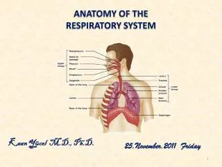
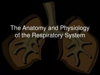
Structural Plan of the Respiratory System • Structure determined by respiratory system functions of air distributor and gas exchanger—supplying oxygen and removing carbon dioxide from cells (Figure 26-1) • Alveoli—sacs that serve as gas exchangers; all other parts of respiratory system serve as air distributors • The respiratory system also warms, filters, and humidifies air • Respiratory organs involved in speech, homeostasis of body pH, and olfaction 2
Structural Plan of the Respiratory System • The respiratory system is divided into two structural divisions • Upper respiratory tract—the organs are located outside the thorax and consist of the nose, nasopharynx, oropharynx, laryngopharynx, and larynx • Lower respiratory tract—the organs are located within the thorax and consist of the trachea, the bronchial tree, and the lungs • Accessory structures include the oral cavity, rib cage, and diaphragm 4
Upper Respiratory Tract • Nose • Structure of the nose—external portion consists of a bony and cartilaginous frame covered by skin containing sebaceous glands • The two nasal bones meet and are surrounded by the frontal bone to form the root • The nose is surrounded by the maxilla (Figure 26-2) 5
Upper Respiratory Tract • Nose (cont) • Internal portion of the nose (nasal cavity) lies over the roof of the mouth, separated by the palatine bones • Cleft palate—condition in which the palatine bones fail to unite completely and only partially separate the nose and the mouth, thereby producing difficulty swallowing • Cribriform plate—separates the roof of the nose from the cranial cavity • Septum—separates the nasal cavity into right and left cavities; it consists of four structures: the perpendicular plate of the ethmoid bone, the vomer bone, the vomeronasal cartilages, and the septal nasal cartilage 7
Upper Respiratory Tract • Nose (cont) • Each nasal cavity is divided into three passageways: superior, middle, and inferior meatuses (Figure 26-3) • Anterior (external) nares—external openings to the nasal cavities; open into the vestibule • Sequence of airflow through the nose into the pharynx—anterior nares to the vestibule to all three meatuses simultaneously and then to the posterior (internal) nares 8
Upper Respiratory Tract • Nose (cont) • Nasal mucosa • Air passes over respiratory mucosa, which contains a rich blood supply (Figure 26-4) • Olfactory epithelium—special sensory membrane containing many olfactory nerve cells and a rich lymphatic plexus 10
Upper Respiratory Tract • Nose (cont) • Paranasal sinuses • Four pairs of air-containing spaces that open or drain into the nasal cavity • Each is lined with respiratory mucosa (Figure 26-5) 12
Upper Respiratory Tract • Nose (cont) • Functions of the nose • Provides a passageway for air traveling to and from the lungs • Filters the air, aids speech, and makes possible the sense of smell 14
Upper Respiratory Tract • Pharynx (throat) • Structure of pharynx • Tubelike structure extending from the base of the skull to the esophagus • Made of muscle and divided into three parts (Figure 26-3)—nasopharynx, oropharynx, and laryngopharynx 15
Upper Respiratory Tract • Pharynx (cont) • Pharyngeal tonsils • Located in the nasopharynx • Called adenoids when they become enlarged • Oropharynx contains two pair of organs—the palatine tonsils (most commonly removed in tonsillectomy) and the lingual tonsils (rarely removed) • Functions of the pharynx—pathway for the respiratory and digestive tracts 16
Upper Respiratory Tract • Larynx (Figures 26-6 and 26-7) • Location of larynx—positioned between the root of the tongue and the upper end of the trachea • Structure of larynx • Consists of cartilages attached to each other by muscle • Lined by a ciliated mucous membrane, which forms two pairs of folds (Figure 26-8)— vestibular folds (false vocal folds) and vocal folds 17
Upper Respiratory Tract • Larynx (cont) • Cartilages (framework) of the larynx—formed by nine cartilages • Single laryngeal cartilages—the three largest cartilages: the thyroid cartilage, the epiglottis, and the cricoid cartilages • Paired laryngeal cartilages—three pairs of smaller cartilages: the arytenoid, the corniculate, and the cuneiform cartilages 21
Upper Respiratory Tract • Larynx (cont) • Muscles of the larynx • Intrinsic muscles both insert and originate within the larynx • Extrinsic muscles insert in the larynx but originate on some other structure • Functions of the larynx—forms part of the airway to the lungs and produces the voice 22
Lower Respiratory Tract • Trachea—often called “windpipe” (Figure 26-10) • Structure of trachea • Extends from the larynx to the primary bronchi • Wall composed of (outer) adventitia, (middle) smooth muscle and C-shaped cartilage rings, (inner) respiratory mucosa; posterior wall is very elastic (Figure 26-11) • Incomplete rings and posterior elasticity allows esophagus to expand into trachea during swallowing • Functions of trachea—furnishes part of the open airway to the lungs; obstruction causes death 23
Lower Respiratory Tract • Bronchi and alveoli • Structure of bronchi • Lower end of the trachea divides into two primary bronchi, one on the right and one on the left • Primary bronchi enter the lung and divide into secondary bronchi, which branch into bronchioles and eventually divide into alveolar ducts and alveoli • 23 levels of branching (Figure 26-12) 26
Lower Respiratory Tract • Bronchi and alveoli (cont) • Structure of alveoli—the primary gas exchange structures • Respiratory membrane—the barrier between which gases are exchanged by alveolar air and blood (Figure 26-15) • Respiratory membrane consists of the alveolar epithelium, the capillary endothelium, and their joined basement membranes • Surfactant—a component of the fluid coating the respiratory membrane that reduces surface tension; produced by type II cells 28
Lower Respiratory Tract • Bronchi and alveoli (cont) • Functions of bronchi and alveoli • Distribute air to the lung’s interior; 23 levels of branching are optimal for oxygen transfer to the blood • Mucus blanket cleans the airways as it is moved upward by the ciliary escalator 30
Lower Respiratory Tract • Lungs • Structure of the lungs—cone-shaped organs extending from the diaphragm to above the clavicles (Figure 26-17) • Hilum—slit on the lung’s medial surface where the primary bronchi and pulmonary blood vessels enter • Base—the inferior surface of the lung that rests on the diaphragm • Costal surface—lies against the ribs 31
Lower Respiratory Tract • Structure of the lungs (cont) • Left lung is divided into two lobes—superior and inferior • Right lung is divided into three lobes—superior, middle, and inferior • Lobes are further divided into functional units—bronchopulmonary segments • Ten segments in the right lung • Eight segments in the left lung • Functions of the lungs—air distribution and gas exchange 33
Lower Respiratory Tract • Thorax (Figure 26-18) • Structure of the thoracic cavity—three divisions divided by the pleura • Pleural divisions—the part occupied by the lungs • Mediastinum—part occupied by the esophagus, trachea, large blood vessels, and heart • Functions of the thorax—brings about inspiration and expiration 34
Cycle of Life: Respiratory System • Respiration may be affected by developmental defects, age-related structural changes, or loss of function throughout the life cycle • Age-related changes affect lung capacity, make ventilation difficult, or reduce the oxygen- or carbon dioxide–carrying capacity of blood • Respiratory efficiency is reduced in old age as a result of changes in ribs, respiratory muscles, and hemoglobin levels 36